| Applications | AC |
| Concentration | 1 mg/ml |
| Specificity | This peptide is specific for NB600-1384 only. |
| Protein/Peptide Type | Antibody Blocking Peptide |
| Gene | MAP1LC3B |
| Dilutions |
|
| Application Notes | This peptide is useful as a blocking peptide for NB600-1384. For further blocking peptide related protocol, click here. |
| Storage | Store at -80C. Avoid freeze-thaw cycles. |
| Buffer | Peptide dissolved in dH20. Contains no BSA. |
| Preservative | No Preservative |
| Concentration | 1 mg/ml |


![Immunohistochemistry ATG7 Antibody (683906) [Unconjugated]](https://images.novusbio.com/images2/ATG7_MAB6608_Immunohistochemistry_10631.jpg)
![Simple Western ATG7 Antibody (683906) [Unconjugated]](https://images.novusbio.com/images2/ATG7_MAB6608_Simple_Western_16409.jpg)
![Western Blot ATG7 Antibody (683906) [Unconjugated]](https://images.novusbio.com/images2/ATG7_MAB6608_Western_Blot_11614.jpg)
![Western Blot AKT [p Ser473] Antibody [Unconjugated] - Pan Specific](https://images.novusbio.com/images2/Akt3_AF887_Western_Blot_5930.jpg)
![Simple Western AKT [p Ser473] Antibody [Unconjugated] - Pan Specific](https://images.novusbio.com/images2/16332.jpg)
![Intracellular Staining by Flow Cytometry AKT [p Ser473] Antibody [Unconjugated] - Pan Specific](https://images.novusbio.com/images2/Akt3_AF887_Flow_Cytometry_8283.jpg)


Research Areas for LC3B Protein (NB600-1384PEP)Find related products by research area.
|
|
Autophagy and RAS signaling: Clinical implications By Christina Towers, PhD The cellular recycling process known as autophagy is currently being targeted in over 60 clinical trials focused on treating different types of cancer1. To date, the only autophagy-targeted ... Read full blog post. |
|
Methamphetamine with HIV induces mitochondrial dysfunction and neuronal injury through oxidative stress By Jamshed Arslan, Pharm. D., PhD. December 1 is the World AIDS Day. Despite the combination antiretroviral therapy, 10-25% of Human Immunodeficiency Virus (HIV)-positive individuals report neurocognitive impairm... Read full blog post. |
|
Autophagy and Metastasis By Christina Towers, PhD The majority of cancer patients die from metastatic disease at secondary sites. The threshold to undergo metastasis is high. Only a minority of cancer cells acquire invasive phenotypes... Read full blog post. |
|
Optogenetic Control of Mitophagy: AMBRA1 based mitophagy switch By Christina Towers, PhD Mitophagy in the BrainSelective autophagic degradation of damaged mitochondria, known as mitophagy, has been described as a cyto-protective process. Accordingly, defects in mitophagy h... Read full blog post. |
|
Read full blog post. |
|
Read full blog post. |
|
Read full blog post. |
|
How to visualize autophagy by microscopy By Christina Towers, PhD Autophagy is a recycling process that relies on the formation of a unique organelle termed an autophagosome. An elegant way to monitor autophagy is through various microscopy techniques to... Read full blog post. |
|
Read full blog post. |
|
Read full blog post. |

The concentration calculator allows you to quickly calculate the volume, mass or concentration of your vial. Simply enter your mass, volume, or concentration values for your reagent and the calculator will determine the rest.
| Gene Symbol | MAP1LC3B |


![Immunohistochemistry-Paraffin TOR/mTOR [p Ser2448] Antibody](https://images.novusbio.com/images/TOR-mTOR-[p-Ser2448]-Antibody-Immunohistochemistry-Paraffin-NB600-607-img0005.jpg)
![Western Blot TOR/mTOR [p Ser2448] Antibody](https://images.novusbio.com/images/TOR-mTOR-[p-Ser2448]-Antibody-Western-Blot-NB600-607-img0006.jpg)
![Data TOR/mTOR [p Ser2448] Antibody](https://images.novusbio.com/images/TOR-mTOR-[p-Ser2448]-Antibody-N-A-NB600-607-img0008.jpg)
![Immunocytochemistry Caspase-3 Antibody [Unconjugated] - Active](https://images.novusbio.com/images2/Caspase-3_AF835_Immunocytochemistry_6532.jpg)
![Immunohistochemistry Caspase-3 Antibody [Unconjugated] - Active](https://images.novusbio.com/images2/Caspase3_AF835_Immunohistochemistry_22976.jpg)
![Immunocytochemistry Caspase-3 Antibody [Unconjugated] - Active](https://images.novusbio.com/images2/Caspase-3_AF835_Immunocytochemistry_9340.jpg)


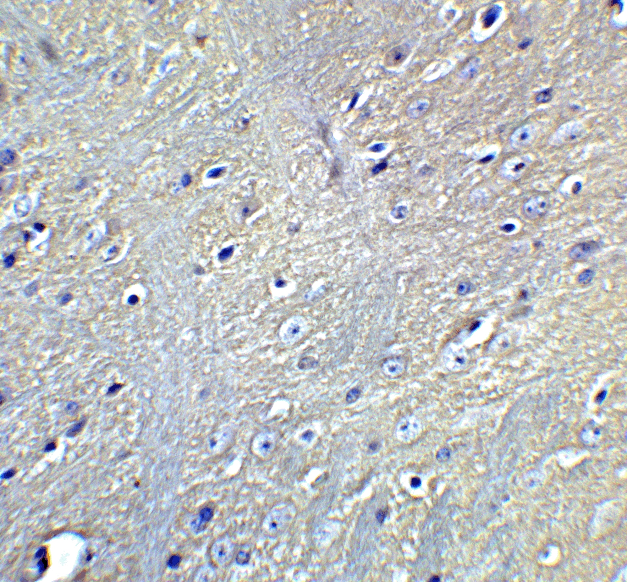
![Analysis using the Biotin conjugate of Rabbit anti-LC3B Antibody [Catalog # NB100-2220]. Staining of brain, cerebral cortex, neurons with cell processes.](https://images.novusbio.com/images/LC3B-Antibody-Immunohistochemistry-NB100-2220-img0053.jpg "Analysis using the Biotin conjugate of Rabbit anti-LC3B Antibody [Catalog # NB100-2220]. Staining of brain, cerebral cortex, neurons with cell processes.")