|
Related Links Exosome Isolation and Detection Exosome Marker Antibodies (CD63, CD81 and more) Exosome Biomarkers for Disease Extracellular Vesicle Flow Cytometry Other Resources Webinar – Flow Analysis of Extracellular Vesicles Liquid Biopsy-Based Biomarkers Whitepaper
|
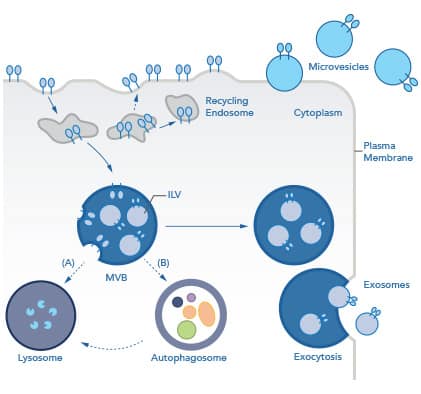
What are exosomes?Once thought to be carriers of unwanted cytosolic waste, extracellular vesicles (EVs) are now understood to be important mediators of intercellular communication under both physiological and pathological conditions. To emphasize the importance of EVs as intercellular messengers, it is worth noting that they have been observed in every Kingdom of life, from the smallest bacteria to every cell in the human body. Exosomes are the smallest EVs and are of particular interest in the tumor microenvironment (TME), where they have been shown to directly mediate angiogenesis, tumor metastasis, and immunosuppression. EVs can be categorized into three main groups based on size, biogenesis, and function:
Request a copy of our Apoptosis, Necroptosis, and Autophagy Poster In place of specific nomenclature relating to biogenesis (e.g. exosome, microvesicle, apoptotic body), the International Society of Extracellular Vesicles (ISEV) recommends referring to EVs based on the following:
For example, if isolating EVs from hypoxia-treated neurons using differential centrifugation and ultrafiltration, they would be “small hypoxic neuronal EVs.” The most stringent way to verify the endosomal nature of exosomes is visually by electron microscopy. Exosome Biogenesis: How are they made?
Exosomes are formed in the endosomal maturation pathway. Briefly, inward budding of endosomes into multivesicular bodies (MVBs) results in the formation of intraluminal vesicles (ILVs). MVBs are destined for one of two fates: degradation or release. MVBs destined for degradation will either (A) fuse directly with the lysosome or (B) fuse with an autophagosome during the process of autophagy. This "amphisome" (MVB plus autophagosome) finally fuses with the lysosome. Alternatively, MVBs destined for release, traffic to the plasma membrane. Upon fusion with the plasma membrane, ILVs are secreted as exosomes via exocytosis.” The rate at which exosomes are secreted varies depending on the cellular source, but certain pathologies, like cancer or hypoxia, can increase the rate of EV release. MVs, in contrast, are vesicles formed directly from the outward protrusions of the plasma membrane and involve cytoskeletal and membrane rearrangement. Both exosomes and MVs are actively secreted from live cells. Apoptotic bodies are unique EVs as they are only formed during programmed cell death, or apoptosis, and are rapidly phagocytosed by patrolling macrophages. Exosome Function: What do they do?EVs are secreted with a diverse array of cargo necessary for intercellular communication, both locally and in distant tissues. This cargo includes genetic information such as messenger RNA (mRNA), microRNA (miRNA), non-coding RNA (ncRNA), as well as lipids and many classes of proteins. Novus Biologicals has an extensive offering of antibodies to “essential” exosome markers, as well as tissue and tumor-specific exosome markers validated for a range of applications including Simple Western and Flow Cytometry. Exosomes and MVs are actively released from many cell types including reticulocytes, lymphocytes, dendritic cells, fibroblasts, endothelial cells, and stem cells as part of a range of dynamic intercellular signaling processes. Roles of Extracellular VesiclesThe Future of ExosomesThe number of applications for exosomes is constantly increasing. In addition to their role as biomarkers for cancers and other diseases, their vesicular nature and wide range of functions make them attractive candidates for therapeutic delivery, regenerative medicine, and vaccine adjuvants.
References:Théry C, Witwer KW, Aikawa E, Alcaraz MJ, Anderson JD, Andriantsitohaina R, et al. Minimal information for studies of extracellular vesicles 2018 (MISEV2018): a position statement of the International Society for Extracellular Vesicles and update of the MISEV2014 guidelines. J Extracell Vesicles 2018;7:. https://doi.org/10.1080/20013078.2018.1535750.
|