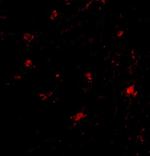
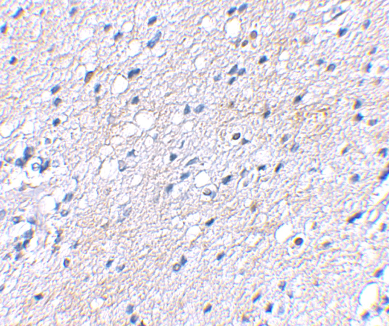
 or double labeled with the G8 and the R&D BAI1 mAbs, or G8 and antibodies to Iba1, NeuN or GFAP. The areas within the boxes of the H&E stained sections are shown at high magnification in the fluorescence photomicrographs. The colors of the fluorescent secondary antibodies are indicated in the unmerged photographs (E, F, H and I). Nuclei were stained with Hoechst dye. Overlap of red and green, when present, appears yellow in merged images (G, J, K, L and M). The G8 and BAI1 mAbs labeled the same subpopulation of cells in the hippocampal formation (box in A; E-G). The Noggin and BAI1 antibodies also bound to the same cells in the glomerular layer of the olfactory bulb (box in B; H-J). G8 did not co-localize with Iba1 (K, from box in A), NeuN (L from lower box in C) or GFAP (M from box in D). Minimal fluorescence was observed with the anti-IgM and anti-IgG (inset in G) or the anti-goat and anti-IgG (inset in J) secondary antibodies. Bar = 270 μM in A-D and 9 μM in E-M. Image collected and cropped by CiteAb from the following open publication (//pubmed.ncbi.nlm.nih.gov/32614850), licensed under a CC-BY license. Not internally tested by R&D Systems.")
| Reactivity | HuSpecies Glossary |
| Applications | IHC |
| Clone | 542515 |
| Clonality | Monoclonal |
| Host | Mouse |
| Conjugate | Unconjugated |
| Concentration | LYOPH |
| Immunogen | Chinese hamster ovary cell line CHO-derived recombinant human BAI1 Met1-Thr879 Accession # O14514 |
| Specificity | Detects human BAI1 in direct ELISAs. In direct ELISAs, no cross-reactivity with recombinant human BAI3 is observed. |
| Source | N/A |
| Isotype | IgG1 |
| Clonality | Monoclonal |
| Host | Mouse |
| Gene | BAI1 |
| Purity Statement | Protein A or G purified from hybridoma culture supernatant |
| Innovator's Reward | Test in a species/application not listed above to receive a full credit towards a future purchase. |
| Dilutions |
|
|
| Publications |
|
| Storage | Use a manual defrost freezer and avoid repeated freeze-thaw cycles.
|
| Buffer | Lyophilized from a 0.2 μm filtered solution in PBS with Trehalose. *Small pack size (SP) is supplied either lyophilized or as a 0.2 µm filtered solution in PBS. |
| Preservative | No Preservative |
| Concentration | LYOPH |
| Reconstitution Instructions | Reconstitute at 0.5 mg/mL in sterile PBS. |


![Western Blot AKT [p Ser473] Antibody [Unconjugated] - Pan Specific](https://images.novusbio.com/images2/Akt3_AF887_Western_Blot_5930.jpg)
![Simple Western AKT [p Ser473] Antibody [Unconjugated] - Pan Specific](https://images.novusbio.com/images2/16332.jpg)
![Intracellular Staining by Flow Cytometry AKT [p Ser473] Antibody [Unconjugated] - Pan Specific](https://images.novusbio.com/images2/Akt3_AF887_Flow_Cytometry_8283.jpg)


![BAI1 Antibody (542515) [Unconjugated]](/sites/all/modules/enterprise-tech/et_datasheets/images/novus_guarantee.png "BAI1 Antibody (542515) [Unconjugated]")
Secondary Antibodies |
Isotype Controls |

The concentration calculator allows you to quickly calculate the volume, mass or concentration of your vial. Simply enter your mass, volume, or concentration values for your reagent and the calculator will determine the rest.


![N/A VEGF [HRP]](https://images.novusbio.com/images2/DATA_VEGF_DVE00_ELISA_871.jpg)
![N/A VEGF [HRP]](https://images.novusbio.com/images2/DATA_VEGF_DVE00_ELISA_872.jpg)
![N/A VEGF [HRP]](https://images.novusbio.com/images2/VEGF_DVE00_ELISA_208.jpg)
![N/A Thrombospondin-1 [HRP]](https://images.novusbio.com/images2/Thrombospondin-1_DTSP10_ELISA_199.jpg)
![N/A Angiopoietin-2 [HRP]](https://images.novusbio.com/images2/Angiopoietin-2_DANG20_ELISA_30.jpg)
![N/A Angiopoietin-2 [HRP]](https://images.novusbio.com/images2/DATA_Angiopoietin2_DANG20_ELISA_574.jpg)
![Flow Cytometry BAI3 Antibody (409633) [Unconjugated]](https://images.novusbio.com/images2/BAI3_MAB39651_Flow_Cytometry_23463.jpg)
![Flow Cytometry BAI3 Antibody (409633) [Unconjugated]](https://images.novusbio.com/images2/BAI3_MAB39651_Flow_Cytometry_23464.jpg)
![N/A Thrombospondin-2 [HRP]](https://images.novusbio.com/images2/Thrombospondin-2_DTSP20_ELISA_200.jpg)
![N/A Thrombospondin-2 [HRP]](https://images.novusbio.com/images2/DATA_Thrombospondin2_DTSP20_ELISA_860.jpg)
![N/A Thrombospondin-2 [HRP]](https://images.novusbio.com/images2/DATA_Thrombospondin2_DTSP20_ELISA_861.jpg)


![N/A IL-10 [Biotin]](https://images.novusbio.com/images2/DATA_IL10_DY417_ELISA_2014.jpg)
