Research of Leukokeratosis, Hereditary Mucosal has been linked to Melanocytic Nevus, Mouth Diseases, Mouth Neoplasms, Leukoplakia, Oral, Hamartoma. The study of Leukokeratosis, Hereditary Mucosal has been mentioned in research publications which can be found using our bioinformatics tool below. Researched pathways related to Leukokeratosis, Hereditary Mucosal include Keratinization, Localization, Response To Antibiotic, Cell Differentiation, Pigmentation. These pathways complement our catalog of research reagents for the study of Leukokeratosis, Hereditary Mucosal including antibodies and ELISA kits against INTERMEDIATE FILAMENT, KERATIN-4, K9, MOTTLED, CARCINOEMBRYONIC ANTIGEN.
Top Research Reagents
We have 2814 products for the study of Leukokeratosis, Hereditary Mucosal that can be applied to Flow Cytometry, Immunocytochemistry/ Immunofluorescence, Immunohistochemistry, Western Blot from our catalog of antibodies and ELISA kits.
Leukokeratosis, Hereditary Mucosal is also known as Hereditary Mucosal Leukokeratosis, Hereditary White Sponge Naevus, White Sponge Naevus, White Sponge Nevus.
![Western Blot: Cytokeratin 4 Antibody (6H9R6) [NBP3-15243] - Western blot analysis of extracts of various cell lines, using Cytokeratin 4 (KRT4) Rabbit mAb (NBP3-15243) at 1:1000 dilution. Secondary antibody: HRP Goat Anti-Rabbit IgG (H+L) at 1:10000 dilution. Lysates/proteins: 25ug per lane. Blocking buffer: 3% nonfat dry milk in TBST. Detection: ECL Basic Kit. Exposure time: 30s.](https://images.novusbio.com/fullsize/Cytokeratin-4-Antibody-6H9R6-Western-Blot-NBP3-15243-img0004.jpg)
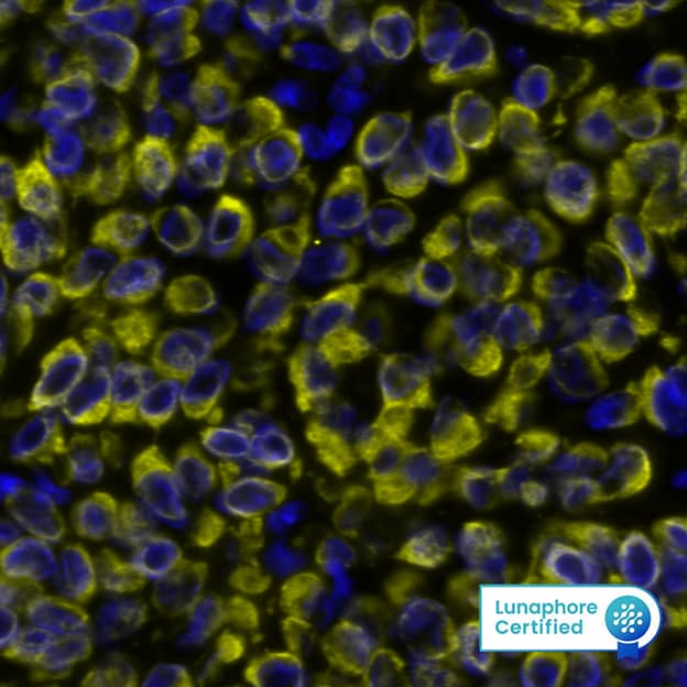
![Immunohistochemistry: Cytokeratin 4 Antibody (6H9R6) [NBP3-15243] - Immunofluorescence analysis of mouse large intestine using Cytokeratin 4 (KRT4) Rabbit mAb (NBP3-15243) at dilution of 1:100 (40x lens). Blue: DAPI for nuclear staining.](https://images.novusbio.com/fullsize/Cytokeratin-4-Antibody-6H9R6-Immunohistochemistry-NBP3-15243-img0003.jpg)
![Western Blot: Cytokeratin 13 Antibody (5M6B10) [NBP3-15261] - Western blot analysis of extracts of various cell lines, using Cytokeratin 13 (KRT13) Rabbit mAb (NBP3-15261) at 1:1000 dilution. Secondary antibody: HRP Goat Anti-Rabbit IgG (H+L) at 1:10000 dilution. Lysates/proteins: 25ug per lane. Blocking buffer: 3% nonfat dry milk in TBST. Detection: ECL Basic Kit. Exposure time: 1s.](https://images.novusbio.com/fullsize/Cytokeratin-13-Antibody-5M6B10-Western-Blot-NBP3-15261-img0005.jpg)
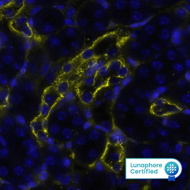
![Immunocytochemistry/Immunofluorescence: Cytokeratin 13 Antibody (5M6B10) [NBP3-15261] - Immunofluorescence analysis of human cervix cancer using Cytokeratin 13 (KRT13) Rabbit mAb (NBP3-15261) at dilution of 1:100 (40x lens). Blue: DAPI for nuclear staining.](https://images.novusbio.com/fullsize/Cytokeratin-13-Antibody-5M6B10-Immunocytochemistry-Immunofluorescence-NBP3-15261-img0003.jpg)
![Western Blot: Cytokeratin 2 Antibody (9T3Q2) [NBP3-16139] - Western blot analysis of extracts of A-431 cells, using Cytokeratin 2 antibody (NBP3-16139) at 1:1000 dilution. Secondary antibody: HRP Goat Anti-Rabbit IgG (H+L) at 1:10000 dilution. Lysates/proteins: 25ug per lane. Blocking buffer: 3% nonfat dry milk in TBST. Detection: ECL Basic Kit. Exposure time: 180s.](https://images.novusbio.com/fullsize/Cytokeratin-2-Antibody-9T3Q2-Western-Blot-NBP3-16139-img0005.jpg)
![Immunohistochemistry: Cytokeratin 2 Antibody (9T3Q2) [NBP3-16139] - Immunofluorescence analysis of mouse skin using Cytokeratin 2 Rabbit mAb (NBP3-16139) at dilution of 1:100 (40x lens). Blue: DAPI for nuclear staining.](https://images.novusbio.com/fullsize/Cytokeratin-2-Antibody-9T3Q2-Immunohistochemistry-NBP3-16139-img0004.jpg)
![ELISA: Human Neuropeptide S/NPS ELISA Kit (Colorimetric) [NBP3-39144] - Standard Curve Reference](https://images.novusbio.com/fullsize/nbp3-39144_human-neuropeptide-s-nps-elisa-kit-colorimetric-211020241536231.png)
![Western Blot: GDI1 Antibody [NBP1-31528] - Sample (30 ug of whole cell lysate) A: Hep G2 10% SDS PAGE; antibody diluted at 1:1000.](https://images.novusbio.com/fullsize/GDI1-Antibody-Western-Blot-NBP1-31528-img0003.jpg)
![Immunohistochemistry-Paraffin: GDI1 Antibody [NBP1-31528] - Sample: Paraffin-embedded mouse lung. GDI1 antibody dilution: 1:500.](https://images.novusbio.com/fullsize/GDI1-Antibody-Immunohistochemistry-Paraffin-NBP1-31528-img0006.jpg)
![Western Blot: Kv6.1 Antibody [NBP1-81572] - Lane 1: NIH-3T3 cell lysate (Mouse embryonic fibroblast cells) Lane 2: NBT-II cell lysate (Rat Wistar bladder tumour cells)](https://images.novusbio.com/fullsize/Kv6.1-Antibody-Western-Blot-NBP1-81572-img0007.jpg)
![Immunocytochemistry/Immunofluorescence: Kv6.1 Antibody [NBP1-81572] - Staining of human cell line U-2 OS shows localization to vesicles. Antibody staining is shown in green.](https://images.novusbio.com/fullsize/Kv6.1-Antibody-Immunocytochemistry-Immunofluorescence-NBP1-81572-img0008.jpg)
![Immunocytochemistry/Immunofluorescence: Plectin Antibody [NBP1-84020] - Plectin antibody in indirect immunofluorescence staining of human tumor cells using a green-fluorescent secondary antibody. Nuclei stained with DAPI (blue). Image from verified customer review.](https://images.novusbio.com/fullsize/Plectin-Antibody-Immunocytochemistry-Immunofluorescence-NBP1-84020-img0017.jpg)
![Immunohistochemistry-Paraffin: Plectin Antibody [NBP1-84020] - Staining of human liver shows no positivity in hepatocytes as expected.](https://images.novusbio.com/fullsize/Plectin-Antibody-Immunohistochemistry-Paraffin-NBP1-84020-img0016.jpg)
![Western Blot: MTSS1 Antibody [NBP2-24716] - Analysis of MTSS1 in human testis lysate in the 1) absence and 2) presence of immunizing peptide, 3) mouse and 4) rat testis lysate using NBP2-24716 at 2 ug/ml, 4 ug/ml and 2 ug/ml, respectively.](https://images.novusbio.com/fullsize/MTSS1-Antibody-Western-Blot-NBP2-24716-img0005.jpg)
![Immunohistochemistry-Paraffin: MTSS1 Antibody [NBP2-24716] - Analysis of human colon using this antibody at 10 ug/ml.](https://images.novusbio.com/fullsize/MTSS1-Antibody-Immunohistochemistry-Paraffin-NBP2-24716-img0004.jpg)
![Immunohistochemistry-Paraffin: Cytokeratin 17 Antibody [NBP2-38536] - Analysis in human urinary bladder and liver tissues using NBP2-38536 antibody. Corresponding Cytokeratin 17 RNA-seq data are presented for the same tissues.](https://images.novusbio.com/fullsize/Cytokeratin-17-Antibody-Immunohistochemistry-Paraffin-NBP2-38536-img0012.jpg)
![Western Blot: Cytokeratin 17 Antibody [NBP2-38536] - Analysis in human cell lines HeLa and PC-3. Corresponding RNA-seq data are presented for the same cell lines. Loading control: Anti-HSP90B1.](https://images.novusbio.com/fullsize/Cytokeratin-17-Antibody-Western-Blot-NBP2-38536-img0011.jpg)
![Immunocytochemistry/Immunofluorescence: Cytokeratin 14 Antibody (LL002) [NBP2-34270] - Immunofluorescence Analysis of A549 cells labeling CK14 with Cytokeratin 14 Antibody (LL002) followed by Goat anti-Mouse IgG-CF488 (Green). The nuclear counterstain is Red Dot (Red).](https://images.novusbio.com/fullsize/Cytokeratin-14-Antibody-LL002-Immunocytochemistry-Immunofluorescence-NBP2-34270-img0010.jpg)
![Immunohistochemistry-Paraffin: Cytokeratin 14 Antibody (LL002) [NBP2-34270] - Formalin-fixed, paraffin-embedded human Prostate Carcinoma stained with Cytokeratin 14 Antibody (LL002).](https://images.novusbio.com/fullsize/Cytokeratin-14-Antibody-LL002-Immunohistochemistry-Paraffin-NBP2-34270-img0011.jpg)
![Western Blot: Cytokeratin 10 Antibody (3C2F5) [NBP2-61736] - Analysis using KRT10 mAb against HEK293 (1) and KRT10 (AA: 345-454)-hIgGFc transfected HEK293 (2) cell lysate.](https://images.novusbio.com/fullsize/Cytokeratin-10-Antibody-3C2F5-Western-Blot-NBP2-61736-img0006.jpg)
![Immunohistochemistry-Paraffin: Cytokeratin 10 Antibody (3C2F5) [NBP2-61736] - Analysis of esophageal cancer tissues using KRT10 mouse mAb with DAB staining.](https://images.novusbio.com/fullsize/Cytokeratin-10-Antibody-3C2F5-Immunohistochemistry-NBP2-61736-img0004.jpg)
![Western Blot: Cytokeratin 5 Antibody [NBP2-61931] - Total protein from human A431 cells and rat PC12 cells was separated on a 7.5% gel by SDS-PAGE, transferred to PVDF membrane and blocked in 5% non-fat milk in TBST. The membrane was probed with 2.0 ug/ml anti-Cytokeratin 5 in 5% non-fat milk in TBST and detected with an anti-rabbit HRP secondary antibody using chemiluminescence.](https://images.novusbio.com/fullsize/Cytokeratin-5-Antibody-Western-Blot-NBP2-61931-img0002.jpg)
![Immunocytochemistry/Immunofluorescence: Cytokeratin 5 Antibody [NBP2-61931] - A431 cells were fixed for 10 minutes using 10% formalin and then permeabilized for 5 minutes using 1X PBS + 0.1% Triton X-100. The cells were incubated with anti-Cytokeratin 5 at 5 ug/ml overnight at 4C and detected with an anti-rabbit DyLight 488 (Green) at a 1:500 dilution. Alpha tubulin (DM1A) NB100-690 was used as a co-stain at a 1:1000 dilution and detected with an anti-mouse DyLight 550 (Red) at a 1:500 dilution. Nuclei were counterstained with DAPI (Blue). Cells were imaged using a 40X objective.](https://images.novusbio.com/fullsize/Cytokeratin-5-Antibody-Immunocytochemistry-Immunofluorescence-NBP2-61931-img0001.jpg)
![Western Blot: Cytokeratin 16 Antibody (SC52-09) [NBP2-67559] - Analysis of Cytokeratin 16 on human skin lysates using anti-Cytokeratin 16 antibody at 1/1,000 dilution.](https://images.novusbio.com/fullsize/Cytokeratin-16-Antibody-SC52-09-Western-Blot-NBP2-67559-img0008.jpg)
![Immunocytochemistry/Immunofluorescence: Cytokeratin 16 Antibody (SC52-09) [NBP2-67559] - Staining Cytokeratin 16 in HepG2 cells (green). The nuclear counter stain is DAPI (blue). Cells were fixed in paraformaldehyde, permeabilised with 0.25% Triton X100/PBS.](https://images.novusbio.com/fullsize/Cytokeratin-16-Antibody-SC52-09-Immunocytochemistry-Immunofluorescence-NBP2-67559-img0004.jpg)
![Western Blot: KCNN3 Antibody [NBP2-85137] - Host: Rabbit. Target Name: KCNN3. Sample Tissue: Human THP-1 Whole Cell. Antibody Dilution: 1ug/ml](https://images.novusbio.com/fullsize/KCNN3-Antibody-Western-Blot-NBP2-85137-img0004.jpg)
![Immunohistochemistry: KCNN3 Antibody [NBP2-85137] - Sample Type: Rhesus macaque spinal cord. Primary Antibody Dilution: 1:300. Secondary Antibody: Donkey anti Rabbit 488. Secondary Antibody Dilution: 1:500. Color/Signal Descriptions: Green: KCNN3. Gene Name: KCNN3. Submitted by: Timur Mavlyutov, Ph. D., Department of Pharmacology, University of Wisconsin Medical School](https://images.novusbio.com/fullsize/KCNN3-Antibody-Immunohistochemistry-NBP2-85137-img0006.jpg)
