Apoptosis is a form of programmed cell death which is mediated by cysteine proteases called caspases. It is an essential phenomenon in the maintenance of homeostasis and growth of tissues, and it also plays a critical role in immune response. The cytomorphological alterations and the key features of apoptosis are listed below:

Process |
Active, physiological or pathophysiological |
Induction stimuli |
Oxidative stress, death receptor ligands, chemotherapy |
Morphological changes |
Nuclear pyknosis, membrane blebbing, generation of apoptotic bodies |
Molecular changes |
Cleavage of caspases and PARP, DNA fragmentation |
Clearance |
Apoptotic bodies phagocytosed by neighboring cells & macrophages |
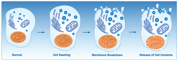
Necroptosis, a programmed necrosis, is a type of cell death which emerges as a backup mechanism when apoptosis is non-functional either genetically or pathogenically. It involves the release of intracellular "danger signals" which results in considerable inflammation. The cytomorphological alterations and the key features of necroptosis are listed below:

Process |
Mostly passive, always pathological |
Induction stimuli |
Viral or chemical exposure, radiation, endogenous or pathological factors |
Morphological changes |
Swelling of cells and organelles, loss of membrane integrity |
Molecular changes |
Acidosis, random DNA degradation, release of cellular proteins |
Clearance |
Necrotic cells ingested by macrophages, significant inflammation |
Autophagy refers to a heterogeneous group of cell signaling pathways which enables eukaryotic cells to deliver cytosolic components to the autophagosomes-lysosomes for degradation, to recycle nutrients, and to survive during starvation. The cytomorphological alterations and the key features of autophagy are listed below:

Process |
Active, physiological or pathophysiological |
Induction stimuli |
Starvation, hypoxia, chemotherapy, growth factor deprivation |
Morphological changes |
Vacuolization, mass degradation of organelles & proteins |
Molecular changes |
LC3I lipidation to LC3II, p62/SQSTM1 degradation, lysosomal activity |
Clearance |
Cells gets cannibalized & the contents recycle for survival of the tissue |
To learn more about these signaling pathways as well as useful tips and protocols to improve your apoptosis assay, request or download your complimentary copy of
| Get Poster: Apoptosis, Necroptosis & Autophagy |
Explore reagents for in your research area: