![Western Blot: PKC iota Antibody [NBP1-84959] - Analysis in human cell line A-431.](http://images.novusbio.com/fullsize/PKC-iota-Antibody-Western-Blot-NBP1-84959-img0008.jpg "Western Blot: PKC iota Antibody [NBP1-84959] - Analysis in human cell line A-431.")
| Reactivity | Hu, Mu, RtSpecies Glossary |
| Applications | WB, ICC/IF, IHC |
| Clonality | Polyclonal |
| Host | Rabbit |
| Conjugate | Unconjugated |
| Immunogen | This antibody was developed against Recombinant Protein corresponding to amino acids: INCKLLVHKKCHKLVTIECGRHSLPQEPVMPMDQSSMHSDHAQTVIPYNPSSHESLDQVGEEKEAMNTRESGKASSSLGLQDFDLLRV |
| Predicted Species | Mouse (93%), Rat (95%). Backed by our 100% Guarantee. |
| Isotype | IgG |
| Clonality | Polyclonal |
| Host | Rabbit |
| Gene | PRKCI |
| Purity | Immunogen affinity purified |
| Innovator's Reward | Test in a species/application not listed above to receive a full credit towards a future purchase. |
| Dilutions |
|
||
| Application Notes | For IHC-Paraffin, HIER pH 6 retrieval is recommended. ICC/IF Fixation Permeabilization: Use PFA/Triton X-100. |
||
| Control Peptide |
|
||
| Publications |
|
| Storage | Store at 4C short term. Aliquot and store at -20C long term. Avoid freeze-thaw cycles. |
| Buffer | PBS (pH 7.2) and 40% Glycerol |
| Preservative | 0.02% Sodium Azide |
| Purity | Immunogen affinity purified |


Secondary Antibodies |
Isotype Controls |
Research Areas for PKC iota Antibody (NBP1-84959)Find related products by research area.
|

The concentration calculator allows you to quickly calculate the volume, mass or concentration of your vial. Simply enter your mass, volume, or concentration values for your reagent and the calculator will determine the rest.
| Gene Symbol | PRKCI |


![Immunocytochemistry/Immunofluorescence: PKC iota Antibody [NBP1-84959] - Staining of human cell line U-251 MG shows localization to cytosol. Antibody staining is shown in green.](http://images.novusbio.com/fullsize/PKC-iota-Antibody-Immunocytochemistry-Immunofluorescence-NBP1-84959-img0007.jpg "Immunocytochemistry/Immunofluorescence: PKC iota Antibody [NBP1-84959] - Staining of human cell line U-251 MG shows localization to cytosol. Antibody staining is shown in green.")
![Immunohistochemistry-Paraffin: PKC iota Antibody [NBP1-84959] - Staining of human Skeletal muscle shows very weak cytoplasmic positivity in myocytes.](http://images.novusbio.com/fullsize/PKC-iota-Antibody-Immunohistochemistry-Paraffin-NBP1-84959-img0014.jpg "Immunohistochemistry-Paraffin: PKC iota Antibody [NBP1-84959] - Staining of human Skeletal muscle shows very weak cytoplasmic positivity in myocytes.")
![Immunohistochemistry-Paraffin: PKC iota Antibody [NBP1-84959] - Staining of human stomach shows strong membranous and cytoplasmic positivity in glandular cells.](http://images.novusbio.com/fullsize/PKC-iota-Antibody-Immunohistochemistry-Paraffin-NBP1-84959-img0009.jpg "Immunohistochemistry-Paraffin: PKC iota Antibody [NBP1-84959] - Staining of human stomach shows strong membranous and cytoplasmic positivity in glandular cells.")
![Immunohistochemistry-Paraffin: PKC iota Antibody [NBP1-84959] - Staining of human fallopian tube shows strong membranous and cytoplasmic positivity in glandular cells.](http://images.novusbio.com/fullsize/PKC-iota-Antibody-Immunohistochemistry-Paraffin-NBP1-84959-img0010.jpg "Immunohistochemistry-Paraffin: PKC iota Antibody [NBP1-84959] - Staining of human fallopian tube shows strong membranous and cytoplasmic positivity in glandular cells.")
![Immunohistochemistry-Paraffin: PKC iota Antibody [NBP1-84959] - Staining of human rectum shows moderate membranous and cytoplasmic positivity in glandular cells.](http://images.novusbio.com/fullsize/PKC-iota-Antibody-Immunohistochemistry-Paraffin-NBP1-84959-img0011.jpg "Immunohistochemistry-Paraffin: PKC iota Antibody [NBP1-84959] - Staining of human rectum shows moderate membranous and cytoplasmic positivity in glandular cells.")
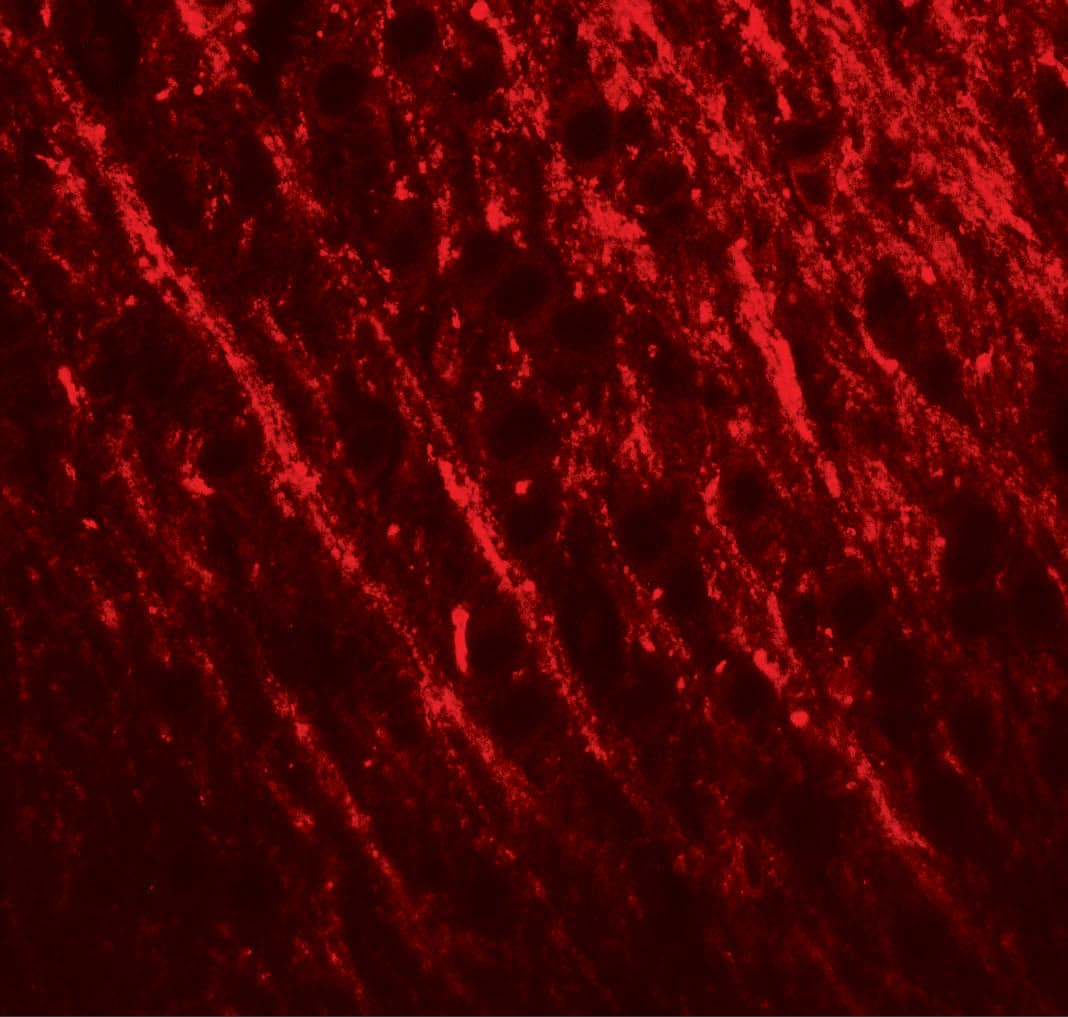
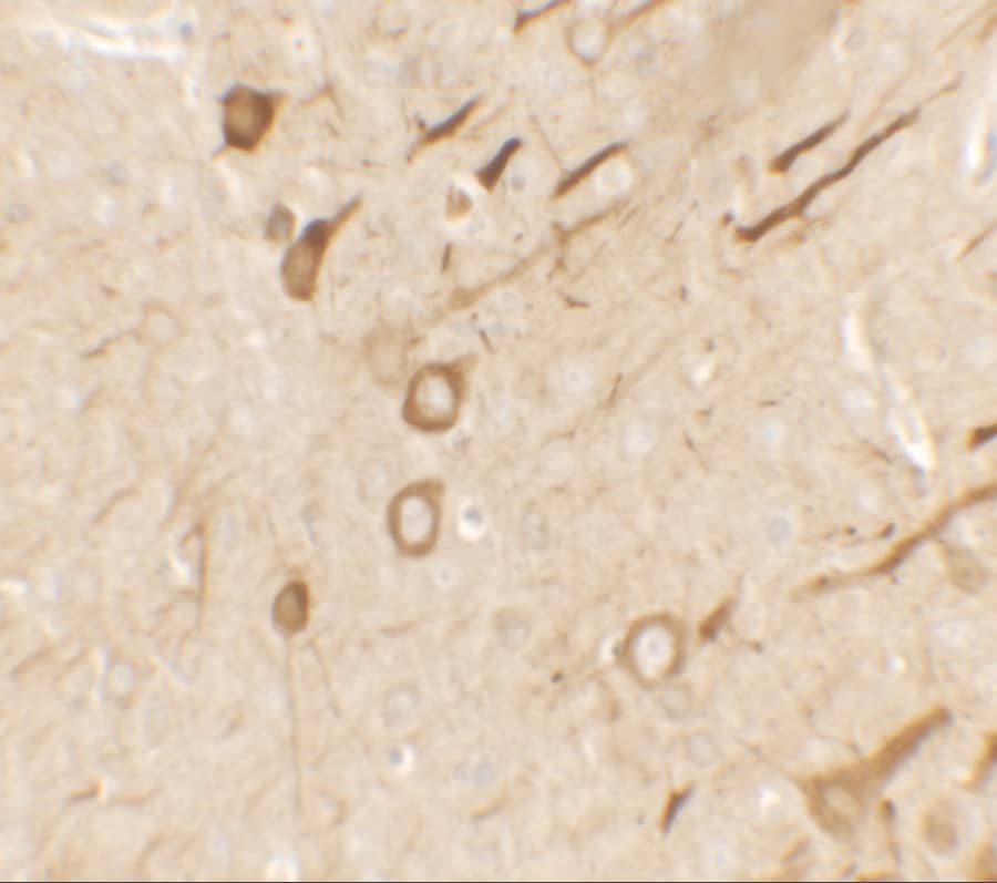
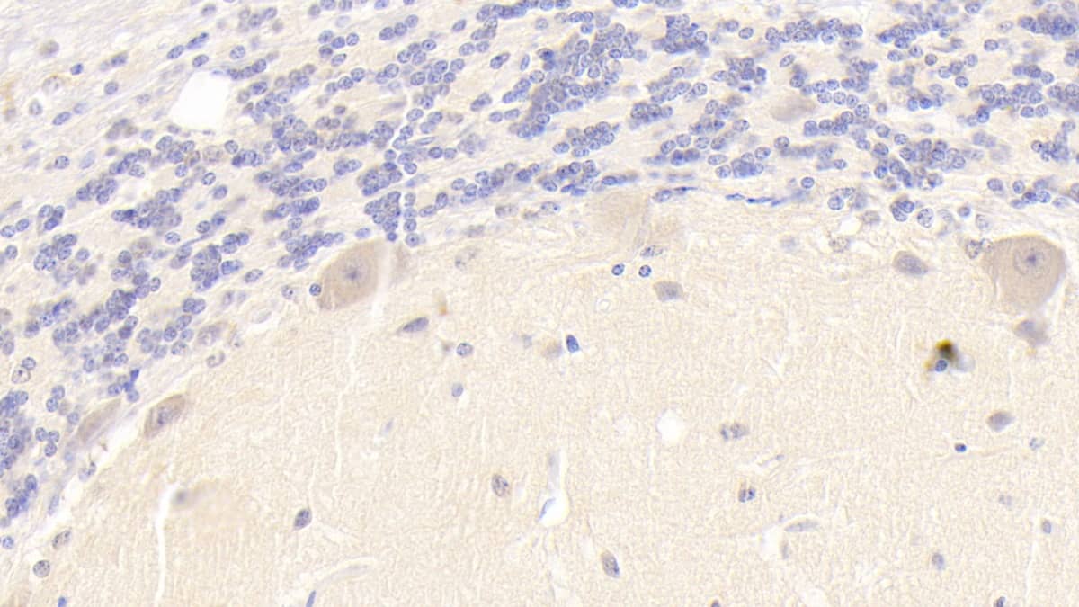
![Immunohistochemistry-Paraffin: PKC iota Antibody [NBP1-84959] - Staining of human cerebral cortex shows strong ctyoplasmic positivity in neurons.](http://images.novusbio.com/fullsize/PKC-iota-Antibody-Immunohistochemistry-Paraffin-NBP1-84959-img0012.jpg "Immunohistochemistry-Paraffin: PKC iota Antibody [NBP1-84959] - Staining of human cerebral cortex shows strong ctyoplasmic positivity in neurons.")
![Immunohistochemistry-Paraffin: PKC iota Antibody [NBP1-84959] - Staining of human skeletal muscle shows very weak cytoplasmic positivity in myocytes.](http://images.novusbio.com/fullsize/PKC-iota-Antibody-Immunohistochemistry-Paraffin-NBP1-84959-img0013.jpg "Immunohistochemistry-Paraffin: PKC iota Antibody [NBP1-84959] - Staining of human skeletal muscle shows very weak cytoplasmic positivity in myocytes.")


![PXR/NR1I2 Antibody (H4417) [Unconjugated]](/sites/all/modules/enterprise-tech/et_datasheets/images/novus_guarantee.png "PXR/NR1I2 Antibody (H4417) [Unconjugated]")

![N/A TIMP-1 [HRP]](https://images.novusbio.com/images2/DATA_TIMP1_DTM100_ELISA_847.jpg)
![N/A TIMP-1 [HRP]](https://images.novusbio.com/images2/TIMP-1_DTM100_ELISA_194.jpg)


![SDS-PAGE TNF-alpha [Unconjugated]](https://images.novusbio.com/images2/TNF-alpha_210-TA_256.jpg)
![Bioactivity TNF-alpha [Unconjugated]](https://images.novusbio.com/images2/TNFalpha_210TA_1658.jpg)
![SEC-MALS TNF-alpha [Unconjugated]](https://images.novusbio.com/images/210-ta_recombinant-human-tnf-alpha-protein-sec-mals-35202312244..jpg)
![Simple Western 14-3-3 zeta Antibody (818515) [Unconjugated]](https://images.novusbio.com/images2/1433_zeta_MAB2669_Simple_Western_16526.jpg)
![Western Blot 14-3-3 zeta Antibody (818515) [Unconjugated]](https://images.novusbio.com/images2/14-3-3_zeta_MAB2669_Western_Blot_12700.jpg)
![Immunohistochemistry 14-3-3 zeta Antibody (818515) [Unconjugated]](https://images.novusbio.com/images2/14-3-3_zeta_MAB2669_Immunohistochemistry_12730.jpg)
![Immunohistochemistry BTK [p Tyr223] Antibody - BSA Free](https://images.novusbio.com/images/BTK-[p-Tyr223]-Antibody-Immunohistochemistry-NBP1-78295-img0002.jpg)
![Western Blot BTK [p Tyr223] Antibody - BSA Free](https://images.novusbio.com/images/BTK-[p-Tyr223]-Antibody-Western-Blot-NBP1-78295-img0004.jpg)
![Western Blot: Goat anti-Rabbit IgG (H+L) Secondary Antibody [HRP] [NB7160] - Western blot showing vemurafenib treatment in BRAFV600E CRC cells inhibits fission mediator DRP1 with no significant effect on fusion proteins (Mfn1 & 2) using MFN-1 antibody (NBP1-51841) and corresponding secondary antibody, goat anti-rabbit IgG-HRP (NB7160). Image collected and cropped by CiteAb from the following publication (https://pubmed.ncbi.nlm.nih.gov/33738242).](https://images.novusbio.com/images/Goat-anti-Rabbit-IgG-H+L-Secondary-Antibody-HRP-Western-Blot-NB7160-img0001.jpg "Western Blot: Goat anti-Rabbit IgG (H+L) Secondary Antibody [HRP] [NB7160] - Western blot showing vemurafenib treatment in BRAFV600E CRC cells inhibits fission mediator DRP1 with no significant effect on fusion proteins (Mfn1 & 2) using MFN-1 antibody (NBP1-51841) and corresponding secondary antibody, goat anti-rabbit IgG-HRP (NB7160). Image collected and cropped by CiteAb from the following publication (https://pubmed.ncbi.nlm.nih.gov/33738242).")
 using a 1:1000 dilution of HRP-conjugated Anti-Rabbit IgG Secondary Antibody (Catalog # HAF008). This experiment was conducted under reducing conditions and using Immunoblot Buffer Group 1.")
![Flow Cytometry: Rabbit IgG Isotype Control [NBP2-24891] - Intracellular FACS analysis of mouse TLR6 polyclonal antibody (red), rabbit isotype control (green), RAW cells alone (shaded). Two micrograms of antibodies were used. Goat anti-rabbit FITC (Novus, 20302) was used as secondary.](https://images.novusbio.com/images/Rabbit--Mouse-IgG-Isotype-Control-Flow-Cytometry-NBP2-24891-img0001.jpg "Flow Cytometry: Rabbit IgG Isotype Control [NBP2-24891] - Intracellular FACS analysis of mouse TLR6 polyclonal antibody (red), rabbit isotype control (green), RAW cells alone (shaded). Two micrograms of antibodies were used. Goat anti-rabbit FITC (Novus, 20302) was used as secondary.")