
| Reactivity | All-NASpecies Glossary |
| Applications | WB, ELISA, IHC, KD, KO |
| Clonality | Polyclonal |
| Host | Goat |
| Conjugate | HRP |
| Conjugate | Catalog # | Availability | Size | Price |
|---|---|---|---|---|
| Alkaline Phosphatase | NB600-1502 | |||
| Biotin | NB100-1678 | |||
| DyLight 488 | NBP1-69969 | |||
| FITC | NB100-1771 | |||
| FITC | NB100-1771SS | |||
| Rhodamine | NBP1-97327-500ug | |||
| Texas Red | NB120-6660 | |||
| Unconjugated | NB100-1770 | |||
| Description | For extended storage aliquot contents and freeze at -20C or below. Avoid cycles of freezing and thawing. Centrifuge product if not completely clear after standing at room This antibody was prepared from monospecific antiserum by immunoaffinity chromatography using Green Fluorescent Protein (Aequorea victoria) coupled to agarose beads followed by solid phase adsorption(s) to remove any unwanted reactivities. Assay by immunoelectrophoresis resulted in a single precipitin arc against anti-Goat Serum, anti-Peroxidase and purified and partially purified Green Fluorescent Protein (Aequorea victoria) |
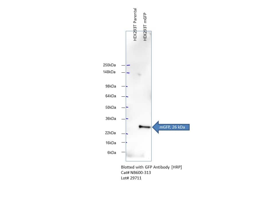
| Immunogen | The immunogen is a Green Fluorescent Protein (GFP) fusion protein corresponding to the full length amino acid sequence (246aa) derived from the jellyfish Aequorea victoria. (Uniprot: P42212) |
| Specificity | No reaction was observed against Human, Mouse and Rat Serum Proteins. |
| Isotype | IgG |
| Clonality | Polyclonal |
| Host | Goat |
| Purity | Immunogen affinity purified |
| Innovator's Reward | Test in a species/application not listed above to receive a full credit towards a future purchase. |
| Dilutions |
|
|
| Application Notes | This product is designed to detect GFP and its variants. Anti-GFP Peroxidase conjugated antibody has been tested by ELISA to detect GFP by ELISA (sandwich or capture) for the direct binding of antigen and recognizes wild type, recombinant and enhanced forms of GFP and by western blot. Biotin conjugated polyclonal anti-GFP used in a sandwich ELISA is well suited to titrate GFP in solution using this antibody in combination with monoclonal anti-GFP using either form of the antibody as the capture or detection antibody. However, use the monoclonal form only for the detection of wild type or recombinant GFP as this form does not sufficiently detect 'enhanced' GFP. The detection antibody is typically conjugated to biotin and subsequently reacted with streptavidin conjugated HRP. Fluorochrome conjugated polyclonal anti-GFP can be used to detect GFP by immunofluorescence microscopy in prokaryotic (E.coli) and eukaryotic (CHO cells) expression systems and can detect GFP containing inserts. Significant amplification of signal is achieved using fluorochrome conjugated polyclonal anti-GFP relative to the fluorescence of GFP alone. For immunoblotting use either alkaline phosphatase or peroxidase conjugated polyclonal anti-GFP to detect GFP or GFP containing proteins on western blots. Optimal titers for applications should be determined by the researcher. NOTE: Do NOT add Sodium Azide (it inhibits HRP irreversibly). |
|
| Readout System | ||
| Reviewed Applications |
|
|
| Publications |
|
| Storage | Store at 4C short term. Aliquot and store at -20C long term. Avoid freeze-thaw cycles. |
| Buffer | 0.02 M Potassium Phosphate, 0.15 M Sodium Chloride, pH 7.2, 10 mg/mL Bovine Serum Albumin (BSA) - Immunoglobulin and Protease free |
| Preservative | 0.01% Gentamicin Sulfate |
| Purity | Immunogen affinity purified |
| Images | Ratings | Applications | Species | Date | Details | ||||||||
|---|---|---|---|---|---|---|---|---|---|---|---|---|---|
Enlarge |
reviewed by:
Verified Customer |
WB | Human | 05/01/2019 |
Summary
|
||||||||
|
reviewed by:
Gummuluru |
WB | Human | 01/20/2012 |
Summary
|
Secondary Antibodies |
Isotype Controls |
Research Areas for GFP Antibody (NB600-313)Find related products by research area.
|
|
Successful Transplantation of Friedreich Ataxia Induced Pluripotent Stem Cell (iPSC)-Derived Sensory Neurons in Dorsal Root Ganglia of Adult Rodents Jamshed Arslan, Pharm D, PhD The dorsal root ganglia (DRG) are a collection of cell bodies of sensory nerves carrying sensory information – including nociception, mechanoreception and proprioception – from periphera... Read full blog post. |
|
Autophagy and RAS signaling: Clinical implications By Christina Towers, PhD The cellular recycling process known as autophagy is currently being targeted in over 60 clinical trials focused on treating different types of cancer1. To date, the only autophagy-targeted ... Read full blog post. |
|
Neurovascular signaling for repair enhances brain metastasis By Jamshed Arslan, Pharm. D., PhD. Stroke is a leading cause of mortality and morbidity worldwide. Cellular players – neurons, astrocytes, endothelial and stromal cells – involved in post-stroke repair t... Read full blog post. |
|
Read full blog post. |
|
How to visualize autophagy by microscopy By Christina Towers, PhD Autophagy is a recycling process that relies on the formation of a unique organelle termed an autophagosome. An elegant way to monitor autophagy is through various microscopy techniques to... Read full blog post. |
|
Toll-like receptors in the intestinal epithelial cells By Jamshed Arslan, Pharm. D., PhD. Toll-like receptors (TLRs) are microbe-sensing proteins that act as first responders to danger signals. TLRs help the intestinal epithelial cells (IECs) recognize commensal bacteria ... Read full blog post. |
|
Read full blog post. |
|
Animal Models to Study Autophagy By Christina Towers, PhD What is autophagy?Autophagy is the catabolic process that degrades cytoplasmic material via the lysosome. The process of macroautophagy was originally characterized in yeast, where the... Read full blog post. |
|
The use of a GFP antibody for research applications in transgenic C. elegans, GFP tagged yeast and porcine model GFP, or green fluorescent protein, is a chemiluminescent protein derived from Aequorea jellyfish that was first discovered by Osamu Shimomura. It was soon after established that the emission spectra of GFP was right around 509nm, or the ultraviol... Read full blog post. |
|
GFP - Be Green! Green fluorescence protein (GFP) is a 27KD protein derived from the jellyfish Aquorea victoria that emits a green light (emission peak at a wavelength of 509 nm) when excited by blue light (excitation peak at a wavelength of 395 nm). GFP is a highly v... Read full blog post. |

The concentration calculator allows you to quickly calculate the volume, mass or concentration of your vial. Simply enter your mass, volume, or concentration values for your reagent and the calculator will determine the rest.
5 | |
4 | |
3 | |
2 | |
1 |
| Verified Customer 05/01/2019 |
||
| Application: | WB | |
| Species: | Human |
| Gummuluru 01/20/2012 |
||
| Application: | WB | |
| Species: | Human |


: none")

 using a 1:1000 dilution of HRP-conjugated Anti-Goat IgG Secondary Antibody (Catalog # HAF017). This experiment was conducted under reducing conditions and using Immunoblot Buffer Group 1.")
