
| Submit your blog on Hair Follicle Development to be featured! |
| Submit your event on Hair Follicle Development to be featured! |
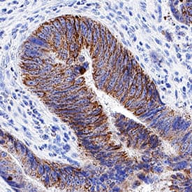
Mouse Monoclonal
Applications IHC
|
|
![Western Blot: LEF1 Antibody (3H5) [H00051176-M01] - LEF1 monoclonal antibody (M01), clone 3H5 Analysis of LEF1 expression in MES-SA/Dx5.](https://images.novusbio.com/fullsize/LEF1-Antibody-3H5-Western-Blot-H00051176-M01-img0009.jpg)
![Western Blot: LEF1 Antibody (3H5) [H00051176-M01] - Analysis of LEF1 over-expressed 293 cell line, cotransfected with LEF1 Validated Chimera RNAi ( Cat # H00051176-R01V ) (Lane 2) or non-transfected control (Lane 1). Blot probed with LEF1 monoclonal antibody (M01), clone 3H5 (Cat # H00051176-M01 ). GAPDH ( 36.1 kDa ) used as specificity and loading control.](https://images.novusbio.com/fullsize/LEF1-Antibody-3H5-Western-Blot-H00051176-M01-img0006.jpg)
Mouse Monoclonal
Species Human, Mouse
Applications WB, ELISA, ICC/IF
| 3 Publications |
|
![Western Blot: SRPR alpha Antibody [H00006734-B02P] - Analysis of SRPR expression in human pancreas.](https://images.novusbio.com/fullsize/SRPR-alpha-Antibody-Western-Blot-H00006734-B02P-img0002.jpg)
![Immunocytochemistry/Immunofluorescence: SRPR alpha Antibody [H00006734-B02P] - Analysis of purified antibody to SRPR on HeLa cell. (antibody concentration 10 ug/ml)](https://images.novusbio.com/fullsize/SRPR-alpha-Antibody-Immunocytochemistry-Immunofluorescence-H00006734-B02P-img0001.jpg)
Mouse Polyclonal
Species Human
Applications WB, ICC/IF
| 1 Publication |
|
![Immunocytochemistry: SGK3 Antibody - BSA Free [NB100-59014] - Immunohistochemical analysis of brain tissue, senile plaques caused by Alzheimer's.](https://images.novusbio.com/fullsize/SGK3-Antibody---BSA-Free-Immunocytochemistry-NB100-59014-img0004.jpg)
![Immunohistochemistry-Paraffin: SGK3 Antibody - BSA Free [NB100-59014] - Anti-SGK3 antibody IHC of human kidney. Immunohistochemistry of formalin-fixed, paraffin-embedded tissue after heat-induced antigen retrieval.](https://images.novusbio.com/fullsize/SGK3-Antibody---BSA-Free-Immunohistochemistry-Paraffin-NB100-59014-img0010.jpg)
Rabbit Polyclonal
Species Human
Applications ELISA, ICC/IF, IHC
|
|
Goat Polyclonal
Species Mouse
Applications WB, Block
| 6 Publications |
|


Goat Polyclonal
Species Human
Applications WB, IHC, ICC
| 8 Publications |
|


Rat Monoclonal
Species Human, Mouse
Applications WB, IHC
| 10 Publications |
|


Goat Polyclonal
Species Human
Applications WB, Simple Western, Flow
| 30 Publications |
|
Species Human
Applications Bind
| 3 Publications |
|


Species Mouse
Applications BA
| 66 Publications |
|


Species Human
Applications BA
| 3 Reviews 808 Publications |
|


Species Human
Applications BA, BA
| 505 Publications |
|


Species Human
Applications BA
| 795 Publications |
|
![Immunohistochemistry-Paraffin: Desmoplakin Antibody [NBP2-48836] - Staining in human skin and skeletal muscle tissues using anti-DSP antibody. Corresponding DSP RNA-seq data are presented for the same tissues.](https://images.novusbio.com/fullsize/Desmoplakin-Antibody-Immunohistochemistry-Paraffin-NBP2-48836-img0005.jpg)
![Immunohistochemistry-Paraffin: Desmoplakin Antibody [NBP2-48836] - Staining of human skeletal muscle shows low expression as expected.](https://images.novusbio.com/fullsize/Desmoplakin-Antibody-Immunohistochemistry-Paraffin-NBP2-48836-img0003.jpg)
Rabbit Polyclonal
Species Human, Canine
Applications ICC/IF, IHC, IHC-P
| 2 Publications |
|
![Knockdown Validated: GLI-1 Antibody - BSA Free [NB600-600] - GLI-1 Antibody [NB600-600] - Inhibition of Hh signaling in CAFs blocks its effects on lymphangiogenesis in vivo and in vitro. (E) IHC staining for Ki-67, -SMA, Gli-1 and LYVE1 in tumour sections from mice in the two groups. Human CAFs promote lymphangiogenesis in ovarian cancer via the Hh-VEGF-C signaling axis. Image collected and cropped by CiteAb from the following publication (//pubmed.ncbi.nlm.nih.gov/31012844/) licensed under a CC-BY license.](https://images.novusbio.com/fullsize/GLI-1-Antibody-Immunohistochemistry-Paraffin-NB600-600-img0005.jpg)
![Immunohistochemistry-Paraffin: GLI-1 Antibody [NB600-600] - Analysis of a FFPE mouse brain section using GLI-1 antibody at 1:600 dilution.](https://images.novusbio.com/fullsize/GLI-1-Antibody-Immunohistochemistry-Paraffin-NB600-600-img0003.jpg)
Rabbit Polyclonal
Species Human, Mouse, Canine
Applications WB, ELISA, IB
| 1 Review 55 Publications |
|
![Western Blot: Isoleucyl tRNA synthetase Antibody [NBP3-13037] - Isoleucyl tRNA synthetase antibody detects Isoleucyl tRNA synthetase protein by western blot analysis. Various whole cell extracts (30 ug) were separated by 5% SDS-PAGE, and the membrane was blotted with Isoleucyl tRNA synthetase antibody (NBP3-13037) diluted at 1:4000.](https://images.novusbio.com/fullsize/Isoleucyl-tRNA-synthetase-Antibody-Western-Blot-NBP3-13037-img0001.jpg)
![Immunohistochemistry-Paraffin: Isoleucyl tRNA synthetase Antibody [NBP3-13037] - Rat brain. Isoleucyl tRNA synthetase antibody (NBP3-13037) diluted at 1:500. Antigen Retrieval: Citrate buffer, pH 6.0, 15 min.](https://images.novusbio.com/fullsize/Isoleucyl-tRNA-synthetase-Antibody-Immunohistochemistry-Paraffin-NBP3-13037-img0002.jpg)
Rabbit Polyclonal
Species Human, Rat
Applications WB, IHC, IHC-P
|
|
