| Submit your blog on Petit Mal Status to be featured! |
| Submit your event on Petit Mal Status to be featured! |
![Western Blot: EPB42 Antibody (2G12) [H00002038-M01] - Analysis of EPB42 expression in transfected 293T cell line by EPB42 monoclonal antibody (M01), clone 2G12.Lane 1: EPB42 transfected lysate(69.5 KDa).Lane 2: Non-transfected lysate.](https://images.novusbio.com/fullsize/EPB42-Antibody-2G12-Western-Blot-H00002038-M01-img0005.jpg)
![Immunocytochemistry/Immunofluorescence: EPB42 Antibody (2G12) [H00002038-M01] - Analysis of monoclonal antibody to EPB42 on HeLa cell . Antibody concentration 10 ug/ml.](https://images.novusbio.com/fullsize/EPB42-Antibody-2G12-Immunocytochemistry-Immunofluorescence-H00002038-M01-img0003.jpg)
Mouse Monoclonal
Species Human
Applications WB, ELISA, ICC/IF
|
|
![Western Blot: Enolase 2/Neuron-specific Enolase Antibody [NB110-58870] - Analysis of different tissue and cell lysates using rabbit pAb to neuron specific enolase (NSE), NB110-58870, dilution 1:5,000 in green: [1] protein standard (red), [2] rat brain, [3] rat spinal cord, [4] mouse brain, [5] mouse spinal cord, [6] NIH-3T3, [7] HEK293, [8] HeLa, [9] SH-SY5Y, and [10] C6 cells. A single band at about 47kDa corresponds to the NSE protein, seen only in extracts containing neurons ro neuronal lineage cells.](https://images.novusbio.com/fullsize/Enolase-2-Neuron-specific-Enolase-Antibody-Western-Blot-NB110-58870-img0007.jpg)
![Immunocytochemistry/Immunofluorescence: Enolase 2/Neuron-specific Enolase Antibody [NB110-58870] - Analysis of mixed cortical neuron-glial cell culture from E20 rat stained with rabbit pAb to neuron specific enolase (NSE), NB110-58870, dilution 1:500 in red, and costained with chicken pAb to GFAP, dilution 1:5,000 in green. The blue is Hoechst staining of nuclear DNA. the NSE antibody labels protein expressed in neuronal cells, while the GFAP antibody stains intermediate filaments in astrocytic and certain other glial cells.](https://images.novusbio.com/fullsize/Enolase-2-Neuron-specific-Enolase-Antibody-Immunocytochemistry-Immunofluorescence-NB110-58870-img0006.jpg)
Rabbit Polyclonal
Species Human, Mouse, Rat
Applications WB, Simple Western, ICC/IF
| 2 Reviews 5 Publications |
|
![Western Blot: Pancreatic Amylase Alpha Antibody (6D4) [NBP1-47659] - Pancreatic Amylase Alpha Antibody (6D4) HEK293T cells were transfected with the pCMV6-ENTRY control (Left lane) or pCMV6-ENTRY Pancreatic Amylase(Right lane) cDNA for 48 hrs and lysed. Equivalent amounts of cell lysates (5 ug per lane) were separated by SDS-PAGE and immunoblotted with anti-Pancreatic Amylase.](https://images.novusbio.com/fullsize/Pancreatic-Amylase-Alpha-Antibody-6D4-Western-Blot-NBP1-47659-img0022.jpg)
![Immunohistochemistry-Paraffin: Pancreatic Amylase Alpha Antibody (6D4) [NBP1-47659] - Pancreatic Amylase Alpha Antibody (6D4) Staining of paraffin-embedded ovary using anti-Pancreatic Amylase mouse monoclonal antibody.](https://images.novusbio.com/fullsize/Pancreatic-Amylase-Alpha-Antibody-6D4-Immunohistochemistry-Paraffin-NBP1-47659-img0021.jpg)
Mouse Monoclonal
Species Human
Applications WB, IHC, IHC-P
| 1 Review |
|
![Flow Cytometry: CD8 Antibody (53-6.7) [NBP1-49045] - CD8 alpha Antibody (53-6.7) [NBP1-49045] - Analysis of lymph nodes by multiple staining.](https://images.novusbio.com/fullsize/CD8-Antibody-53-6-7-Flow-Cytometry-NBP1-49045-img0014.jpg)
![Immunohistochemistry-Paraffin: CD8 Antibody (53-6.7) [NBP1-49045] - CD8 alpha Antibody (53-6.7) [NBP1-49045] - CD8 alpha expression in mouse spleen tissue using anti-CD8 alpha antibody. Image from verified customer review.](https://images.novusbio.com/fullsize/CD8-Antibody-53-6-7-Immunohistochemistry-Paraffin-NBP1-49045-img0004.jpg)
Rat Monoclonal
Species Mouse, Rat
Applications Flow, ICC/IF, IHC
| 3 Reviews 28 Publications |
|
![Western Blot: Mkl1 Antibody [NBP1-88498] - Analysis in U-251MG cells transfected with control siRNA, target specific siRNA probe #1 and #2. Remaining relative intensity is presented. Loading control: Anti-GAPDH.](https://images.novusbio.com/fullsize/Mkl1-Antibody-Western-Blot-NBP1-88498-img0010.jpg)
![Immunocytochemistry/Immunofluorescence: Mkl1 Antibody [NBP1-88498] - Staining of human cell line A-431 shows localization to nucleoplasm. Antibody staining is shown in green.](https://images.novusbio.com/fullsize/Mkl1-Antibody-Immunocytochemistry-Immunofluorescence-NBP1-88498-img0008.jpg)
Rabbit Polyclonal
Species Human, Mouse
Applications WB, ICC/IF, IHC
| 9 Publications |
|
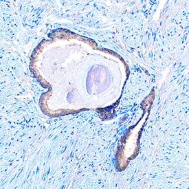
![Immunohistochemistry-Paraffin: Cav2.2 Antibody [NBP1-91744] - Staining of human cerebral cortex shows moderate cytoplasmic positivity in glial cells.](https://images.novusbio.com/fullsize/Cav2.2-Antibody-Immunohistochemistry-Paraffin-NBP1-91744-img0003.jpg)
Rabbit Polyclonal
Species Human
Applications IHC, IHC-P
|
|
![Western Blot: Pallidin Antibody (1H9) [NBP2-01763] - HEK293T cells were transfected with the pCMV6-ENTRY control (Left lane) or pCMV6-ENTRY Pallidin (Right lane) cDNA for 48 hrs and lysed. Equivalent amounts of cell lysates (5 ug per lane) were separated by SDS-PAGE and immunoblotted with anti-Pallidin.](https://images.novusbio.com/fullsize/Pallidin-Antibody-1H9-Western-Blot-NBP2-01763-img0007.jpg)
![Immunohistochemistry-Paraffin: Pallidin Antibody (1H9) [NBP2-01763] - Staining of paraffin-embedded Human tonsil using anti-Pallidin mouse monoclonal antibody.](https://images.novusbio.com/fullsize/Pallidin-Antibody-1H9-Immunohistochemistry-Paraffin-NBP2-01763-img0006.jpg)
Mouse Monoclonal
Species Human, Mouse, Rat
Applications WB, Flow, IHC
|
|
![SDS-Page: PRH1 Protein [NBP2-23368]](https://images.novusbio.com/fullsize/PRH1-Protein-SDS-Page-NBP2-23368-img0001.jpg)
Species Human
Applications PAGE
|
|
![TIRAP (TLR2 and TLR4) Inhibitor Peptide [NBP2-26245] - TLR2 acts through formation of heterodimer complexes with TLR1 or TLR6. HEK 293 endogenously expresses TLR1 and TLR6, so that the TLR2 reporter cell line can response to both Pam3CSK4 (TLR2/TLR1 specific ligand) and MALP-2 (TLR2/TLR6 specific ligand). The TLR2 / TLR4 inhibitor specifically inhibits TLR2/TLR1 receptor complex activity in a dose-response manner but exhibited no or little effect on TLR2/TLR6 receptor complex activity.](https://images.novusbio.com/fullsize/TIRAP-TLR2-and-TLR4-Inhibitor-Peptide-NBP2-26245-img0007.jpg)
![TIRAP (TLR2 and TLR4) Inhibitor Peptide [NBP2-26245] - The TLR4 reporter cell line responds to LPS. The TLR2 / TLR4 inhibitor specifically inhibits TLR4 activation upon LPS stimulation in a dose-response manner.](https://images.novusbio.com/fullsize/TIRAP-TLR2-and-TLR4-Inhibitor-Peptide-NBP2-26245-img0008.jpg)
Species Human, Mouse
Applications Flow, In vitro
| 8 Publications |
|
![Immunohistochemistry: POMC Antibody [NB100-1533] - Representative confocal images of POMC in POMC-transfected WT and Sel1L-/- N2a cells. White arrows point to POMC-containing secretory granules, while yellow arrows point to perinuclear POMC. KDEL marks the ER. Representative data from at least 2 independent experiments are shown. Image collected and cropped by CiteAb from the following publication (jci.org/articles/view/96420), licensed under a CC-BY license.](https://images.novusbio.com/fullsize/POMC-Antibody-Immunohistochemistry-NB100-1533-img0008.jpg)
![Flow Cytometry: POMC Antibody [NB100-1533] - Flow cytometric analysis of paraformaldehyde fixed A431 cells (blue line), permeabilized with 0.5% Triton. Primary incubation 1hr (10 ug/mL) followed by Alexa Fluor 488 secondary antibody (1 ug/mL). IgG control: Unimmunized goat IgG (black line) followed by Alexa Fluor 488 secondary antibody.](https://images.novusbio.com/fullsize/POMC-Antibody-Flow-Cytometry-NB100-1533-img0006.jpg)
Goat Polyclonal
Species Human, Mouse, Rat
Applications WB, Flow, ICC/IF
| 11 Publications |
|

Goat Polyclonal
Species Human, Mouse, Rat
Applications WB, IHC
| 1 Review |
|


Goat Polyclonal
Species Mouse, Rat
Applications WB, Simple Western, IHC
| 7 Publications |
|
Species Human
Applications EnzAct
| 2 Publications |
|

Species Human
Applications BA
| 3 Publications |
|
![Immunohistochemistry-Paraffin: LAMC2 Antibody (CL2980) [NBP2-42388] - Staining in human fallopian tube and liver tissues. Corresponding LAMC2 RNA-seq data are presented for the same tissues.](https://images.novusbio.com/fullsize/LAMC2-Antibody-CL2980-Immunohistochemistry-Paraffin-NBP2-42388-img0023.jpg)
![Western Blot: LAMC2 Antibody (CL2980) [NBP2-42388] - Analysis in A-431 cells transfected with control siRNA, target specific siRNA probe #1 and #2, using Anti-LAMC2 antibody. Remaining relative intensity is presented. Loading control: Anti-GAPDH.](https://images.novusbio.com/fullsize/LAMC2-Antibody-CL2980-Western-Blot-NBP2-42388-img0017.jpg)
Mouse Monoclonal
Species Human
Applications WB, ICC/IF, IHC
| 4 Publications |
|
![Immunocytochemistry/Immunofluorescence: ARSE Antibody [NBP2-56601] - Staining of human cell line Hep G2 shows localization to the Golgi apparatus.](https://images.novusbio.com/fullsize/ARSE-Antibody-Immunocytochemistry-Immunofluorescence-NBP2-56601-img0001.jpg)
Rabbit Polyclonal
Species Human
Applications ICC/IF
|
|
![Western Blot: MAL Antibody (B5-G3) [NBP2-75563] - Analysis of MAL on mouse kidney (1) and mouse lymphatic vessels (2) tissue lysate using anti-MAL antibody at 1/1,000 dilution.](https://images.novusbio.com/fullsize/MAL-Antibody-B5-G3-Western-Blot-NBP2-75563-img0006.jpg)
![Immunohistochemistry-Paraffin: MAL Antibody (B5-G3) [NBP2-75563] - Analysis of paraffin-embedded rat kidney tissue using anti-MAL antibody. Counter stained with hematoxylin.](https://images.novusbio.com/fullsize/MAL-Antibody-B5-G3-Immunohistochemistry-Paraffin-NBP2-75563-img0005.jpg)
Mouse Monoclonal
Species Human, Mouse, Rat
Applications WB, Flow, IHC
|
|
![Immunocytochemistry/Immunofluorescence: SQLE Antibody [NBP2-93808] - Analysis of NIH-3T3 cells using SQLE . Blue: DAPI for nuclear staining.](https://images.novusbio.com/fullsize/SQLE-Antibody-Immunocytochemistry-Immunofluorescence-NBP2-93808-img0002.jpg)
![Immunocytochemistry/Immunofluorescence: SQLE Antibody [NBP2-93808] - Analysis of HeLa cells using SQLE . Blue: DAPI for nuclear staining.](https://images.novusbio.com/fullsize/SQLE-Antibody-Immunocytochemistry-Immunofluorescence-NBP2-93808-img0001.jpg)
Rabbit Polyclonal
Species Human, Mouse, Rat
Applications WB, ICC/IF, IHC
|
|
![Western Blot: SKIP Antibody [NBP2-94173] - Analysis of extracts of Mouse skeletal muscle, using INPP5K antibody at 1:1000 dilution. Secondary antibody: HRP Goat Anti-Rabbit IgG (H+L) at 1:10000 dilution.Lysates/proteins: 25ug per lane. Blocking buffer: 3% nonfat dry milk in TBST.Detection: ECL Basic Kit. Exposure time: 60s.](https://images.novusbio.com/fullsize/SKIP-Antibody-Western-Blot-NBP2-94173-img0003.jpg)
![Immunocytochemistry/Immunofluorescence: SKIP Antibody [NBP2-94173] - Analysis of PC-12 cells using INPP5K Rabbit pAb at dilution of 1:150 (40x lens). Blue: DAPI for nuclear staining.](https://images.novusbio.com/fullsize/SKIP-Antibody-Immunocytochemistry-Immunofluorescence-NBP2-94173-img0007.jpg)
Rabbit Polyclonal
Species Human, Mouse, Rat
Applications WB, ICC/IF
|
|
![SDS-Page: Recombinant Human PRH2 Protein [H00005555-P01] - 12.5% SDS-PAGE Stained with Coomassie Blue.](https://images.novusbio.com/fullsize/qc_test-H00005555-P01-1.jpg)
Species Human
Applications WB, ELISA, PA
|
|
