
| Submit your blog on Mycoplasma Pneumonia to be featured! |
| Submit your event on Mycoplasma Pneumonia to be featured! |
![Western Blot: PTGER2 Antibody (2G10L6) [NBP3-16740] - Analysis of extracts of various cell lines, using PGE Receptor EP2 (PGE Receptor EP2 (PTGER2)) Rabbit mAb (NBP3-16740) at 1:1000 dilution. Secondary antibody: HRP Goat Anti-Rabbit IgG (H+L) at 1:10000 dilution. Lysates/proteins: 25ug per lane. Blocking buffer: 3% nonfat dry milk in TBST. Detection: ECL Basic Kit. Exposure time: 3s.](https://images.novusbio.com/fullsize/PTGER2-Antibody-2G10L6-Western-Blot-NBP3-16740-img0005.jpg)
![Immunocytochemistry/Immunofluorescence: PTGER2 Antibody (2G10L6) [NBP3-16740] - Immunofluorescence analysis of U-2 OS cells using PGE Receptor EP2 (PGE Receptor EP2 (PTGER2)) Rabbit mAb (NBP3-16740) at dilution of 1:100 (40x lens). Blue: DAPI for nuclear staining.](https://images.novusbio.com/fullsize/PTGER2-Antibody-2G10L6-Immunocytochemistry-Immunofluorescence-NBP3-16740-img0004.jpg)
Rabbit Monoclonal
Species Human, Mouse, Rat
Applications WB, ICC/IF, IHC
|
|
Mouse Monoclonal
Applications IHC
|
|
![N/A IL-6 [HRP]](https://images.novusbio.com/fullsize/d6050b_human-il-6-quantikine-elisa-kit-2762023115954.png)
![N/A IL-6 [HRP]](https://images.novusbio.com/fullsize/d6050b_human-il-6-quantikine-elisa-kit-276202312352.jpg)
Species Human
| 766 Publications |
|
![Western Blot: CD77 Synthase Antibody [NBP1-62583] - THP-1 cell lysate, concentration 0.2-1 ug/ml.](https://images.novusbio.com/fullsize/CD77-Synthase-Antibody-Western-Blot-NBP1-62583-img0002.jpg)
Rabbit Polyclonal
Species Human
Applications WB
| 2 Publications |
|
![Immunohistochemistry-Paraffin: DBT Antibody [NBP1-89522] - Analysis in human kidney and pancreas tissues. Corresponding DBT RNA-seq data are presented for the same tissues.](https://images.novusbio.com/fullsize/DBT-Antibody-Immunohistochemistry-Paraffin-NBP1-89522-img0012.jpg)
![Immunohistochemistry-Paraffin: DBT Antibody [NBP1-89522] - Staining of human colon, liver, lymph node and pancreas using Anti-DBT antibody NBP1-89522 (A) shows similar protein distribution across tissues to independent antibody NBP1-85964 (B).](https://images.novusbio.com/fullsize/DBT-Antibody-Immunohistochemistry-Paraffin-NBP1-89522-img0013.jpg)
Rabbit Polyclonal
Species Human, Mouse, Rat
Applications WB, ICC/IF, IHC
|
|
![Immunohistochemistry-Paraffin: COX-2 Antibody [NB100-689] - Formalin fixed paraffin embedded colon carcinoma stained with COX-2 antibody (NB100-689).](https://images.novusbio.com/fullsize/COX-2-Antibody-Immunohistochemistry-Paraffin-NB100-689-img0010.jpg)
![Simple Western: COX-2 Antibody [NB100-689] - Simple Western lane view shows a specific band for COX2 in 1.0 mg/ml of Rat Brain at an antibody concentration of 1:100. This experiment was performed under reducing conditions using the 12-230 kDa separation system. Image provided through customer review.](https://images.novusbio.com/fullsize/COX-2-Antibody-Simple-Western-NB100-689-img0003.jpg)
Rabbit Polyclonal
Species Human, Rat
Applications WB, Simple Western, IHC
| 1 Review 36 Publications |
|
![Immunohistochemistry: POMC Antibody [NB100-1533] - Representative confocal images of POMC in POMC-transfected WT and Sel1L-/- N2a cells. White arrows point to POMC-containing secretory granules, while yellow arrows point to perinuclear POMC. KDEL marks the ER. Representative data from at least 2 independent experiments are shown. Image collected and cropped by CiteAb from the following publication (jci.org/articles/view/96420), licensed under a CC-BY license.](https://images.novusbio.com/fullsize/POMC-Antibody-Immunohistochemistry-NB100-1533-img0008.jpg)
![Flow Cytometry: POMC Antibody [NB100-1533] - Flow cytometric analysis of paraformaldehyde fixed A431 cells (blue line), permeabilized with 0.5% Triton. Primary incubation 1hr (10 ug/mL) followed by Alexa Fluor 488 secondary antibody (1 ug/mL). IgG control: Unimmunized goat IgG (black line) followed by Alexa Fluor 488 secondary antibody.](https://images.novusbio.com/fullsize/POMC-Antibody-Flow-Cytometry-NB100-1533-img0006.jpg)
Goat Polyclonal
Species Human, Mouse, Rat
Applications WB, Flow, ICC/IF
| 10 Publications |
|


Goat Polyclonal
Species Human, Mouse, Rat
Applications WB, Simple Western, ICC
| 3 Reviews 15 Publications |
|
![N/A Erythropoietin/EPO [HRP]](https://images.novusbio.com/fullsize2/DATA_Erythropoietin_MEP00_ELISA_970.jpg)
![N/A Erythropoietin/EPO [HRP]](https://images.novusbio.com/fullsize2/Erythropoietin_MEP00_ELISA_421.jpg)
Species Mouse
Applications ELISA
| 104 Publications |
|


Species Human
Applications BA
| 3 Reviews 808 Publications |
|
![Immunocytochemistry/Immunofluorescence: PTGFR Antibody [NLS1049] - Representative FPr immunofluorescent staining on porcine corpus luteum-microvascular endothelial cells (pCL-MVECs) isolated from the early luteal phase (EL-p) (A), the mid-luteal phase (ML-p). Representative immunostaining of the FPr on non-fixed EL-p pCL-MVECs is shown (E). Image collected and cropped by CiteAb from the following publication (rbej.biomedcentral.com/articles/10.1186/1477-7827-5-31), licensed under a CC-BY license.](https://images.novusbio.com/fullsize/PTGFR-Antibody-Immunocytochemistry-Immunofluorescence-NLS1049-img0007.jpg)
![Immunohistochemistry-Paraffin: PTGFR Antibody [NLS1049] - Analysis of anti-FP / PTGFR antibody with human skin, melanoma.](https://images.novusbio.com/fullsize/PTGFR-Antibody-Immunohistochemistry-Paraffin-NLS1049-img0005.jpg)
Rabbit Polyclonal
Species Human, Porcine, Equine
Applications ICC/IF, IHC, IHC-P
| 2 Publications |
|
![Western Blot: PMEL17/SILV Antibody (HMB45) [NBP2-44520] - Western Blot Analysis of COLO-38 cell lysate using PMEL17/SILV antibody (HMB45).](https://images.novusbio.com/fullsize/PMEL17-SILV-Antibody-HMB45-Western-Blot-NBP2-44520-img0008.jpg)
![Immunohistochemistry: PMEL17/SILV Antibody (HMB45) [NBP2-44520] - PMEL17 (red) was detected in human skin (melanoma) using PMEL17-PE antibody (1:200) in PBS for 1 hour. Nuclei were stained with DAPI (blue). Image from a verified customer review. Image using the PE format of this antibody.](https://images.novusbio.com/fullsize/PMEL17-SILV-Antibody-HMB45-Immunohistochemistry-NBP2-44520-img0007.jpg)
Mouse Monoclonal
Species Human, Canine (Negative), Rat (Negative)
Applications WB, Flow, ICC/IF
| 8 Publications |
|

Mouse Monoclonal
Species Human
Applications IHC
|
|
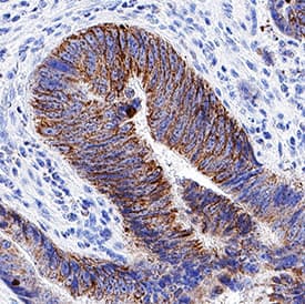
![Immunohistochemistry-Paraffin: PTGER4/EP4 Antibody [NLS3890] - Colon, Carcinoma](https://images.novusbio.com/fullsize/PTGER4-EP4-Antibody-Immunohistochemistry-Paraffin-NLS3890-img0010.jpg)
![Immunohistochemistry-Paraffin: PTGER4/EP4 Antibody [NLS3890] - Analysis of anti-PTGER4 / EP4 antibody with human colon, ganglion at 2 ug/ml.](https://images.novusbio.com/fullsize/PTGER4-EP4-Antibody-Immunohistochemistry-Paraffin-NLS3890-img0003.jpg)
Rabbit Polyclonal
Species Human, Bovine, Monkey
Applications IHC, IHC-P
| 2 Publications |
|


Mouse Monoclonal
Species Human
Applications WB, Flow, CyTOF-ready
|
|
![Western Blot: ACBP Antibody [NBP2-92856] - Analysis of extracts of various cell lines, using DBI antibody at 1:1000 dilution.Secondary antibody: HRP Goat Anti-Rabbit IgG (H+L) at 1:10000 dilution.Lysates/proteins: 25ug per lane. Blocking buffer: 3% nonfat dry milk in TBST.Detection: ECL Basic Kit. Exposure time: 180s.](https://images.novusbio.com/fullsize/ACBP-Antibody-Western-Blot-NBP2-92856-img0005.jpg)
![Immunocytochemistry/Immunofluorescence: ACBP Antibody [NBP2-92856] - Immunofluorescence analysis of U2OS cells using ACBP Rabbit pAb (NBP2-92856) at dilution of 1:100 (40x lens). Blue: DAPI for nuclear staining.](https://images.novusbio.com/fullsize/ACBP-Antibody-Immunocytochemistry-Immunofluorescence-NBP2-92856-img0009.jpg)
Rabbit Polyclonal
Species Human, Mouse, Rat
Applications WB, ICC/IF, IHC
|
|
![Western Blot: SDHA Antibody (GT20710) [NBP3-13522] - Non-transfected (-) and transfected (+) 293T whole cell extracts (30 ug) were separated by 7.5% SDS-PAGE, and the membrane was blotted with SDHA antibody [GT20710] (NBP3-13522) diluted at 1:1000.](https://images.novusbio.com/fullsize/SDHA-Antibody-GT20710-Western-Blot-NBP3-13522-img0009.jpg)
![Immunocytochemistry/Immunofluorescence: SDHA Antibody (GT20710) [NBP3-13522] - SDHA antibody [GT20710] detects SDHA protein at mitochondria by immunofluorescent analysis. Sample: HeLa cells were fixed in 4% paraformaldehyde at RT for 15 min. Green: SDHA protein stained by SDHA antibody [GT20710] (NBP3-13522) diluted at 1:500. Red: Phalloidin, a cytoskeleton marker, diluted at 1:100. Blue: Hoechst 33342 staining.](https://images.novusbio.com/fullsize/SDHA-Antibody-GT20710-Immunocytochemistry-Immunofluorescence-NBP3-13522-img0004.jpg)
Mouse Monoclonal
Species Human, Mouse, Rat
Applications WB, ICC/IF, IHC
|
|
