
| Submit your blog on Keratoderma, Palmoplantar, Epidermolytic to be featured! |
| Submit your event on Keratoderma, Palmoplantar, Epidermolytic to be featured! |
![Western Blot: GDI1 Antibody [NBP1-31528] - Sample (30 ug of whole cell lysate) A: Hep G2 10% SDS PAGE; antibody diluted at 1:1000.](https://images.novusbio.com/fullsize/GDI1-Antibody-Western-Blot-NBP1-31528-img0003.jpg)
![Immunohistochemistry-Paraffin: GDI1 Antibody [NBP1-31528] - Sample: Paraffin-embedded mouse lung. GDI1 antibody dilution: 1:500.](https://images.novusbio.com/fullsize/GDI1-Antibody-Immunohistochemistry-Paraffin-NBP1-31528-img0006.jpg)
Rabbit Polyclonal
Species Human, Mouse
Applications WB, IHC, IHC-P
|
|
![Flow Cytometry: CD8 Antibody (53-6.7) [NBP1-49045] - CD8 alpha Antibody (53-6.7) [NBP1-49045] - Analysis of lymph nodes by multiple staining.](https://images.novusbio.com/fullsize/CD8-Antibody-53-6-7-Flow-Cytometry-NBP1-49045-img0014.jpg)
![Immunohistochemistry-Paraffin: CD8 Antibody (53-6.7) [NBP1-49045] - CD8 alpha Antibody (53-6.7) [NBP1-49045] - CD8 alpha expression in mouse spleen tissue using anti-CD8 alpha antibody. Image from verified customer review.](https://images.novusbio.com/fullsize/CD8-Antibody-53-6-7-Immunohistochemistry-Paraffin-NBP1-49045-img0004.jpg)
Rat Monoclonal
Species Mouse, Rat
Applications Flow, ICC/IF, IHC
| 3 Reviews 28 Publications |
|
![Immunohistochemistry-Paraffin: Filaggrin Antibody [NBP1-87528] - Analysis in human skin and liver tissues. Corresponding Filaggrin RNA-seq data are presented for the same tissues.](https://images.novusbio.com/fullsize/Filaggrin-Antibody-Immunohistochemistry-Paraffin-NBP1-87528-img0017.jpg)
![Immunohistochemistry-Paraffin: Filaggrin Antibody [NBP1-87528] - Staining of human gastrointestinal, liver, skin and testis using Anti-Filaggrin antibody NBP1-87528 (A) shows similar protein distribution across tissues to independent antibody NBP1-87527 (B).](https://images.novusbio.com/fullsize/Filaggrin-Antibody-Immunohistochemistry-Paraffin-NBP1-87528-img0018.jpg)
Rabbit Polyclonal
Species Human
Applications ICC/IF, IHC, IHC-P
| 1 Review 7 Publications |
|
![TIRAP (TLR2 and TLR4) Inhibitor Peptide [NBP2-26245] - TLR2 acts through formation of heterodimer complexes with TLR1 or TLR6. HEK 293 endogenously expresses TLR1 and TLR6, so that the TLR2 reporter cell line can response to both Pam3CSK4 (TLR2/TLR1 specific ligand) and MALP-2 (TLR2/TLR6 specific ligand). The TLR2 / TLR4 inhibitor specifically inhibits TLR2/TLR1 receptor complex activity in a dose-response manner but exhibited no or little effect on TLR2/TLR6 receptor complex activity.](https://images.novusbio.com/fullsize/TIRAP-TLR2-and-TLR4-Inhibitor-Peptide-NBP2-26245-img0007.jpg)
![TIRAP (TLR2 and TLR4) Inhibitor Peptide [NBP2-26245] - The TLR4 reporter cell line responds to LPS. The TLR2 / TLR4 inhibitor specifically inhibits TLR4 activation upon LPS stimulation in a dose-response manner.](https://images.novusbio.com/fullsize/TIRAP-TLR2-and-TLR4-Inhibitor-Peptide-NBP2-26245-img0008.jpg)
Species Human, Mouse
Applications Flow, In vitro
| 8 Publications |
|
Mouse Monoclonal
Species Human, Mouse, Rat
Applications WB, Flow, ICC/IF
| 1 Review 67 Publications |
|
Goat Polyclonal
Species Mouse
Applications WB, IP
| 4 Publications |
|
![Western Blot: Connexin 26/GJB2 Antibody [NBP2-41304] - Western blotting analysis detects Cx26 using R(alpha)Cx26-Ct or R(alpha)Cx26-cl antibodies. (a) R(alpha)Cx26-Ct (A) and R(alpha)Cx26-cl (B) antibodies detected a specific 26kDa band on pig heart tissue lysates (lanes A and B are cropped blots obtained from the same gel). R(alpha)Cx26-cl recognized the 26kDa band on human and rat heart (H) lysates and on rat liver (L) sample (positive control) although two unspecific bands at molecular weights lower than 26kDa were detected. No bands were detected on smooth muscle (SM) tissue lysates (negative control) by R(alpha)Cx26-cl. R(alpha)GAPDH was used as protein loading control for rat tissue samples run on the same gel. Image collected and cropped by CiteAb from the following publication (nature.com/articles/s41598-018-32405-2), licensed under a CC-BY license.](https://images.novusbio.com/fullsize/Connexin-26-GJB2-Antibody-Western-Blot-NBP2-41304-img0005.jpg)
![Immunocytochemistry/Immunofluorescence: Connexin 26/GJB2 Antibody [NBP2-41304] - Cx26 distribution at level of the cardiomyocyte cytoplasm. Confocal laser scanning microscopy: representative images of rat and pig heart sections. Double immunofluorescence analysis for Cx26 (red) and Cx43 (green) show a distinct cell localization of the two Cxs. (a) Three dimensional picture of the maximum intensity projection of the raw images of longitudinal rat heart section treated with R(alpha)Cx26-cl and M(alpha)Cx43. Scale bar: 20um. Image collected and cropped by CiteAb from the following publication (nature.com/articles/s41598-018-32405-2), licensed under a CC-BY license.](https://images.novusbio.com/fullsize/Connexin-26-GJB2-Antibody-Immunocytochemistry-Immunofluorescence-NBP2-41304-img0002.jpg)
Rabbit Polyclonal
Species Human, Rat, Porcine
Applications WB, ELISA, ICC/IF
| 5 Publications |
|
![Immunocytochemistry/Immunofluorescence: Cytokeratin 14 Antibody (LL002) [NBP2-34270] - Immunofluorescence Analysis of A549 cells labeling CK14 with Cytokeratin 14 Antibody (LL002) followed by Goat anti-Mouse IgG-CF488 (Green). The nuclear counterstain is Red Dot (Red).](https://images.novusbio.com/fullsize/Cytokeratin-14-Antibody-LL002-Immunocytochemistry-Immunofluorescence-NBP2-34270-img0010.jpg)
![Immunohistochemistry-Paraffin: Cytokeratin 14 Antibody (LL002) [NBP2-34270] - Formalin-fixed, paraffin-embedded human Prostate Carcinoma stained with Cytokeratin 14 Antibody (LL002).](https://images.novusbio.com/fullsize/Cytokeratin-14-Antibody-LL002-Immunohistochemistry-Paraffin-NBP2-34270-img0011.jpg)
Mouse Monoclonal
Species Human, Mouse, Rat
Applications Simple Western, Flow, ICC/IF
| 4 Publications |
|
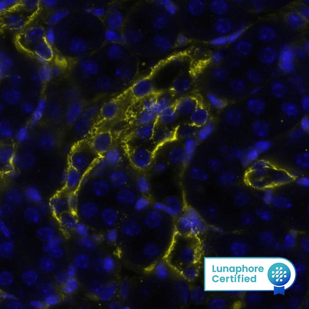
![Immunohistochemistry-Paraffin: Desmoplakin Antibody [NBP2-48836] - Staining in human skin and skeletal muscle tissues using anti-DSP antibody. Corresponding DSP RNA-seq data are presented for the same tissues.](https://images.novusbio.com/fullsize/Desmoplakin-Antibody-Immunohistochemistry-Paraffin-NBP2-48836-img0005.jpg)
![Immunohistochemistry-Paraffin: Desmoplakin Antibody [NBP2-48836] - Staining of human skeletal muscle shows low expression as expected.](https://images.novusbio.com/fullsize/Desmoplakin-Antibody-Immunohistochemistry-Paraffin-NBP2-48836-img0003.jpg)
Rabbit Polyclonal
Species Human, Canine
Applications ICC/IF, IHC, IHC-P
| 2 Publications |
|
![Western Blot: Cytokeratin 10 Antibody (3C2F5) [NBP2-61736] - Analysis using KRT10 mAb against HEK293 (1) and KRT10 (AA: 345-454)-hIgGFc transfected HEK293 (2) cell lysate.](https://images.novusbio.com/fullsize/Cytokeratin-10-Antibody-3C2F5-Western-Blot-NBP2-61736-img0006.jpg)
![Immunohistochemistry-Paraffin: Cytokeratin 10 Antibody (3C2F5) [NBP2-61736] - Analysis of esophageal cancer tissues using KRT10 mouse mAb with DAB staining.](https://images.novusbio.com/fullsize/Cytokeratin-10-Antibody-3C2F5-Immunohistochemistry-NBP2-61736-img0004.jpg)
Mouse Monoclonal
Species Human, Mouse, Rat
Applications WB, ELISA, Flow
| 3 Publications |
|
![Western Blot: Cytokeratin 5 Antibody [NBP2-61931] - Total protein from human A431 cells and rat PC12 cells was separated on a 7.5% gel by SDS-PAGE, transferred to PVDF membrane and blocked in 5% non-fat milk in TBST. The membrane was probed with 2.0 ug/ml anti-Cytokeratin 5 in 5% non-fat milk in TBST and detected with an anti-rabbit HRP secondary antibody using chemiluminescence.](https://images.novusbio.com/fullsize/Cytokeratin-5-Antibody-Western-Blot-NBP2-61931-img0002.jpg)
![Immunocytochemistry/Immunofluorescence: Cytokeratin 5 Antibody [NBP2-61931] - A431 cells were fixed for 10 minutes using 10% formalin and then permeabilized for 5 minutes using 1X PBS + 0.1% Triton X-100. The cells were incubated with anti-Cytokeratin 5 at 5 ug/ml overnight at 4C and detected with an anti-rabbit DyLight 488 (Green) at a 1:500 dilution. Alpha tubulin (DM1A) NB100-690 was used as a co-stain at a 1:1000 dilution and detected with an anti-mouse DyLight 550 (Red) at a 1:500 dilution. Nuclei were counterstained with DAPI (Blue). Cells were imaged using a 40X objective.](https://images.novusbio.com/fullsize/Cytokeratin-5-Antibody-Immunocytochemistry-Immunofluorescence-NBP2-61931-img0001.jpg)
Rabbit Polyclonal
Species Human, Rat
Applications WB, Flow, ICC/IF
| 2 Publications |
|
![Western Blot: Cytokeratin 16 Antibody (SC52-09) [NBP2-67559] - Analysis of Cytokeratin 16 on human skin lysates using anti-Cytokeratin 16 antibody at 1/1,000 dilution.](https://images.novusbio.com/fullsize/Cytokeratin-16-Antibody-SC52-09-Western-Blot-NBP2-67559-img0008.jpg)
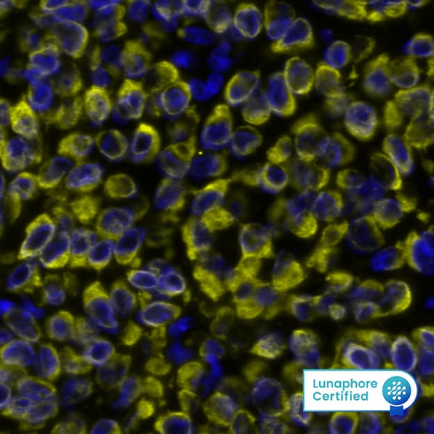
![Immunocytochemistry/Immunofluorescence: Cytokeratin 16 Antibody (SC52-09) [NBP2-67559] - Staining Cytokeratin 16 in HepG2 cells (green). The nuclear counter stain is DAPI (blue). Cells were fixed in paraformaldehyde, permeabilised with 0.25% Triton X100/PBS.](https://images.novusbio.com/fullsize/Cytokeratin-16-Antibody-SC52-09-Immunocytochemistry-Immunofluorescence-NBP2-67559-img0004.jpg)
Rabbit Monoclonal
Species Human
Applications WB, Flow, ICC/IF
|
|
![Western Blot: KRT9 Antibody (SD201-09) [NBP2-67810] - Analysis of KRT9 on A431 cell lysates. Proteins were transferred to a PVDF membrane and blocked with 5% BSA in PBS for 1 hour at room temperature. The primary antibody (1/500) was used in 5% BSA at room temperature for 2 hours. Goat Anti-Rabbit IgG - HRP Secondary Antibody at 1:200,000 dilution was used for 1 hour at room temperature.](https://images.novusbio.com/fullsize/KRT9-Antibody-SD201-09-Western-Blot-NBP2-67810-img0002.jpg)
![Immunocytochemistry/Immunofluorescence: KRT9 Antibody (SD201-09) [NBP2-67810] - Analysis of MCF-7 cells labeling KRT9 (and then blocked with 2% negative goat serum for 15 minutes at room temperature. Cells were probed with the primary antibody KRT9 (red) at 1/200 dilution for overnight at 4 . Goat Anti-Rabbit IgG H&L (iFluor(TM) 594) was used as the secondary antibody at 1/800 dilution. DAPI was used to stain the cell nuclei (blue).](https://images.novusbio.com/fullsize/KRT9-Antibody-SD201-09-Immunocytochemistry-Immunofluorescence-NBP2-67810-img0003.jpg)
Rabbit Monoclonal
Species Human, Mouse, Rat
Applications WB, ICC/IF, IHC
|
|
