| Submit your blog on Epidermolysis Bullosa to be featured! |
| Submit your event on Epidermolysis Bullosa to be featured! |
![Western Blot: NOD2 Antibody (2D9) [NB100-524] - HCMV infection induces NOD2 mRNA and protein in HFFs and U373 cells. E. U373 glioma cells were infected with HCMV Towne strain and levels of NOD1, NOD2 and GAPDH mRNAs were measured by qRT-PCR at indicated time points. F. HFFs were infected with HCMV (Towne) at MOI of 1 PFU/cell and levels of NOD2 protein and B-actin were determined 48 and 72 hpi. G. HFFs were infected with HCMV (Towne) strain at MOI of 0.03 or 3 PFU/cell and levels of NOD2 protein and B-actin were determined at 48 hpi. Quantitative data represent mean values (+/-SD) of triplicate determinations from three independent experiments (*p<0.05, **p<0.01, ***p<0.001, one-way ANOVA test). Image collected and cropped by CiteAb from the following publication (//doi.org/10.1371/journal.pone.0092704.g001) licensed under a CC-BY license.](https://images.novusbio.com/fullsize/NOD2-Antibody-2D9-Western-Blot-NB100-524-img0018.jpg)
![Immunohistochemistry-Frozen: NOD2 Antibody (2D9) [NB100-524] - Overlay of NOD2-DyLight 488 (green) with phase contrast of murine colon. Image from verified customer review.](https://images.novusbio.com/fullsize/NOD2-Antibody-2D9-Immunohistochemistry-Frozen-NB100-524-img0016.jpg)
Mouse Monoclonal
Species Human, Mouse
Applications WB, Flow, ICC/IF
| 1 Review 26 Publications |
|
![Immunocytochemistry/Immunofluorescence: Plectin Antibody [NBP1-84020] - Plectin antibody in indirect immunofluorescence staining of human tumor cells using a green-fluorescent secondary antibody. Nuclei stained with DAPI (blue). Image from verified customer review.](https://images.novusbio.com/fullsize/Plectin-Antibody-Immunocytochemistry-Immunofluorescence-NBP1-84020-img0017.jpg)
![Immunohistochemistry-Paraffin: Plectin Antibody [NBP1-84020] - Staining of human liver shows no positivity in hepatocytes as expected.](https://images.novusbio.com/fullsize/Plectin-Antibody-Immunohistochemistry-Paraffin-NBP1-84020-img0016.jpg)
Rabbit Polyclonal
Species Human
Applications ICC/IF, IHC, IHC-P
| 1 Review |
|
![Immunocytochemistry/Immunofluorescence: Dystonin Antibody [NBP1-89946] - Staining of human cell line A-431 shows localization to nucleus & cytosol. Antibody staining is shown in green.](https://images.novusbio.com/fullsize/Dystonin-Antibody-Immunocytochemistry-Immunofluorescence-NBP1-89946-img0004.jpg)
![Immunohistochemistry-Paraffin: Dystonin Antibody [NBP1-89946] - Staining of human tonsil shows low positivity in non-germinal center cells as expected.](https://images.novusbio.com/fullsize/Dystonin-Antibody-Immunohistochemistry-Paraffin-NBP1-89946-img0008.jpg)
Rabbit Polyclonal
Species Human
Applications ICC/IF, IHC, IHC-P
|
|
![Western Blot: Fibronectin Antibody - BSA Free [NBP1-91258] - VSOP observed in perivascular-restricted spinal cord lesions with intact BBB. Immunostaining for laminin (brown) shows vascular endothelium and glia limitans of a perivascular lesion, along with infiltrating cells and VSOP (blue). Image collected and cropped by CiteAb from the following publication (https://asn.sagepub.com/lookup/doi/10.1042/AN20120081), licensed under a CC-BY license.](https://images.novusbio.com/fullsize/Fibronectin-Antibody---BSA-Free-Western-Blot-NBP1-91258-img0020.jpg)
![Immunocytochemistry/Immunofluorescence: Fibronectin Antibody - BSA Free [NBP1-91258] - NIH3T3 cells were fixed in 4% paraformaldehyde for 10 minutes and permeabilized in 0.05% Triton X-100 in PBS for 5 minutes. The cells were incubated with anti- NBP1-91258 at 1 ug/ml overnight at 4C and detected with an anti-rabbit Dylight 488 (Green) at a 1:1000 dilution for 60 minutes. Nuclei were counterstained with DAPI (Blue). Cells were imaged using a 100X objective and digitally deconvolved.](https://images.novusbio.com/fullsize/Fibronectin-Antibody---BSA-Free-Immunocytochemistry-Immunofluorescence-NBP1-91258-img0021.jpg)
Rabbit Polyclonal
Species Human, Mouse, Rat
Applications WB, Simple Western, ICC/IF
| 11 Reviews 56 Publications |
|
Mouse Monoclonal
Species Human, Mouse, Rat
Applications WB, Flow, ICC/IF
| 1 Review 71 Publications |
|

Goat Polyclonal
Species Mouse
Applications WB, IHC
| 1 Review 16 Publications |
|


Mouse Monoclonal
Species Human
Applications WB, IHC, IP
| 4 Publications |
|


Goat Polyclonal
Species Mouse, Rat
Applications WB, IHC
| 61 Publications |
|

Species Human
Applications BA
| 3 Publications |
|
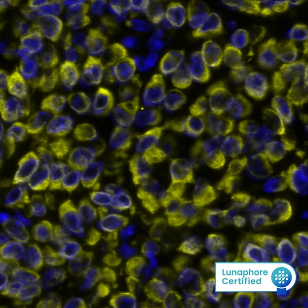
![Immunohistochemistry-Paraffin: LAMC2 Antibody (CL2980) [NBP2-42388] - Staining in human fallopian tube and liver tissues. Corresponding LAMC2 RNA-seq data are presented for the same tissues.](https://images.novusbio.com/fullsize/LAMC2-Antibody-CL2980-Immunohistochemistry-Paraffin-NBP2-42388-img0023.jpg)
![Western Blot: LAMC2 Antibody (CL2980) [NBP2-42388] - Analysis in A-431 cells transfected with control siRNA, target specific siRNA probe #1 and #2, using Anti-LAMC2 antibody. Remaining relative intensity is presented. Loading control: Anti-GAPDH.](https://images.novusbio.com/fullsize/LAMC2-Antibody-CL2980-Western-Blot-NBP2-42388-img0017.jpg)
Mouse Monoclonal
Species Human
Applications WB, ICC/IF, IHC
| 4 Publications |
|
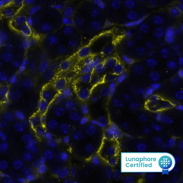
![Immunohistochemistry-Paraffin: Laminin beta 3 Antibody (CL3353) [NBP2-46622] - Analysis in human skin and pancreas tissues using NBP2-46622 antibody. Corresponding LAMB3 RNA-seq data are presented for the same tissues.](https://images.novusbio.com/fullsize/Laminin-beta-3-Antibody-CL3353-Immunohistochemistry-Paraffin-NBP2-46622-img0021.jpg)
![Western Blot: Laminin beta 3 Antibody (CL3353) [NBP2-46622] - Analysis of purified human recombinant Laminin-332, Laminin-421, Laminin-511, Laminin-121 and Laminin-221.](https://images.novusbio.com/fullsize/Laminin-beta-3-Antibody-CL3353-Western-Blot-NBP2-46622-img0018.jpg)
Mouse Monoclonal
Species Human, Rat
Applications WB, ICC/IF, IHC
| 1 Review 2 Publications |
|
![Immunocytochemistry/Immunofluorescence: Cytokeratin 14 Antibody (LL002) [NBP2-34270] - Immunofluorescence Analysis of A549 cells labeling CK14 with Cytokeratin 14 Antibody (LL002) followed by Goat anti-Mouse IgG-CF488 (Green). The nuclear counterstain is Red Dot (Red).](https://images.novusbio.com/fullsize/Cytokeratin-14-Antibody-LL002-Immunocytochemistry-Immunofluorescence-NBP2-34270-img0010.jpg)
![Immunohistochemistry-Paraffin: Cytokeratin 14 Antibody (LL002) [NBP2-34270] - Formalin-fixed, paraffin-embedded human Prostate Carcinoma stained with Cytokeratin 14 Antibody (LL002).](https://images.novusbio.com/fullsize/Cytokeratin-14-Antibody-LL002-Immunohistochemistry-Paraffin-NBP2-34270-img0011.jpg)
Mouse Monoclonal
Species Human, Mouse, Rat
Applications Simple Western, Flow, ICC/IF
| 4 Publications |
|
![Western Blot: Cytokeratin 5 Antibody [NBP2-61931] - Total protein from human A431 cells and rat PC12 cells was separated on a 7.5% gel by SDS-PAGE, transferred to PVDF membrane and blocked in 5% non-fat milk in TBST. The membrane was probed with 2.0 ug/ml anti-Cytokeratin 5 in 5% non-fat milk in TBST and detected with an anti-rabbit HRP secondary antibody using chemiluminescence.](https://images.novusbio.com/fullsize/Cytokeratin-5-Antibody-Western-Blot-NBP2-61931-img0002.jpg)
![Immunocytochemistry/Immunofluorescence: Cytokeratin 5 Antibody [NBP2-61931] - A431 cells were fixed for 10 minutes using 10% formalin and then permeabilized for 5 minutes using 1X PBS + 0.1% Triton X-100. The cells were incubated with anti-Cytokeratin 5 at 5 ug/ml overnight at 4C and detected with an anti-rabbit DyLight 488 (Green) at a 1:500 dilution. Alpha tubulin (DM1A) NB100-690 was used as a co-stain at a 1:1000 dilution and detected with an anti-mouse DyLight 550 (Red) at a 1:500 dilution. Nuclei were counterstained with DAPI (Blue). Cells were imaged using a 40X objective.](https://images.novusbio.com/fullsize/Cytokeratin-5-Antibody-Immunocytochemistry-Immunofluorescence-NBP2-61931-img0001.jpg)
Rabbit Polyclonal
Species Human, Rat
Applications WB, Flow, ICC/IF
| 2 Publications |
|
![Immunocytochemistry/Immunofluorescence: Collagen XVII Antibody (SR46-05) [NBP2-67316] - Staining Collagen XVII in HUVEC cells (green). The nuclear counter stain is DAPI (blue). Cells were fixed in paraformaldehyde, permeabilised with 0.25% Triton X100/PBS.](https://images.novusbio.com/fullsize/Collagen-XVII-Antibody-SR46-05-Immunocytochemistry-Immunofluorescence-NBP2-67316-img0001.jpg)
![Western Blot: Collagen XVII Antibody (SR46-05) [NBP2-67316] - Analysis of Collagen XVII on different lysates with Rabbit anti-Collagen XVII antibody at 1/5,000 dilution.Lane 1: A431 cell lysate (15 ug/Lane)Lane 2: Mouse skin tissue lysate (20 ug/Lane)Lane 3: Rat skin tissue lysate (20 ug/Lane)Predicted band size: 148 kDaObserved band size: 130、180 kDaExposure time: 40 seconds; ECL: K1802;4-20% SDS-PAGE gel.Proteins were transferred to a PVDF membrane and blocked with 5% NFDM/TBST for 1 hour at room temperature. The primary antibody at 1/5,000 dilution was used in 5% NFDM/TBST at room temperature for 2 hours. Goat Anti-Rabbit IgG - HRP Secondary Antibody at 1/50,000 dilution was used for 1 hour at room temperature.](https://images.novusbio.com/fullsize/nbp2-67316_rabbit-monoclonal-collagen-xvii-antibody-sr46-05-western-blot-26720249233..jpg)
Rabbit Monoclonal
Species Human, Mouse, Rat
Applications WB, ICC/IF, IHC
| 1 Publication |
|
![Western Blot: HLA DQ/DR/DP Antibody (HLA-Pan/2967R) - Azide and BSA Free [NBP2-79843] - Western Blot Analysis of Ramos cell lysate using HLA DQ/DR/DP Antibody (HLA-Pan/2967R).](https://images.novusbio.com/fullsize/HLA-DQ-DR-DP-Antibody-HLA-Pan-2967R-Azide-and-BSA-Free-Western-Blot-NBP2-79843-img0003.jpg)
![Immunocytochemistry/Immunofluorescence: HLA DQ/DR/DP Antibody (HLA-Pan/2967R) - Azide and BSA Free [NBP2-79843] - Immunofluorescence staining of PFA-fixed Ramos cells. HLA DQ/DR/DP Recombinant Rabbit Monoclonal Antibody (HLA DQ/DR/DP/2967R) followed by goat anti-rabbit IgG-CF488 (green). Nuclei stained with RedDot.](https://images.novusbio.com/fullsize/HLA-DQ-DR-DP-Antibody-HLA-Pan-2967R-Azide-and-BSA-Free-Immunocytochemistry-Immunofluorescence-NBP2-79843-img0006.jpg)
Rabbit Monoclonal
Species Human
Applications WB, ELISA, Flow
|
|
![Immunocytochemistry/Immunofluorescence: Collagen VII Antibody (LH7.2) [NBP3-07751] - Immunofluorescence Analysis of PFA-fixed HeLa cells labeling Collagen VIIusing Collagen VII Mouse Monoclonal Antibody (LH7.2) followed by goat anti- Mouse IgG-CF488 (Green). The nuclear counterstain is RedDot.](https://images.novusbio.com/fullsize/Collagen-VII-Antibody-LH7-2-Immunocytochemistry-Immunofluorescence-NBP3-07751-img0002.jpg)
![Immunohistochemistry-Paraffin: Collagen VII Antibody (LH7.2) [NBP3-07751] - Formalin-fixed, paraffin-embedded human Skin stained with Collagen VII Mouse Monoclonal Antibody (LH7.2).](https://images.novusbio.com/fullsize/Collagen-VII-Antibody-LH7-2-Immunohistochemistry-Paraffin-NBP3-07751-img0004.jpg)
Mouse Monoclonal
Species Human, Bovine, Equine
Applications WB, ELISA, Flow
|
|
