
| Submit your blog on Eccrine Dermal Cylindroma to be featured! |
| Submit your event on Eccrine Dermal Cylindroma to be featured! |
![Western Blot: Vimentin Antibody [NB300-223] - Analysis of tissue and cell lysates. Antibody at 1:5000 in red. [1] protein standard (red), [2] rat whole brain lysate, [3] HeLa, [4] SH-SY5Y, [5] HEK293, and [6] NIH-3T3 cell lysates. NB300-223 binds to the vimentin protein showing a single band at ~50 kDa. The blot was simultaneously probed with mouse mAb to MAP2C/D, dilution 1:5000 in green, revealing multiple bands around 280 kDa that correspond to full length MAP2A/2B isotypes, and ~70 kDa bands which are MAP2C/D isotypes. MAP2 isotypes are seen only in extracts containing neuronal lineage cells.](https://images.novusbio.com/fullsize/Vimentin-Antibody-Western-Blot-NB300-223-img0002.jpg)
![Immunohistochemistry: Vimentin Antibody [NB300-223] - Attenuated reactive astrocytosis after stroke in Smad1 cKO mice. Enlarged IHC images of boxed areas at the cortical peri-infarct area with the indicated reactive astrocyte markers GFAP, Nestin, and Vimentin. Enlarged images of GFAP IHC highlight the hypertrophic morphology of GFAP+ astrocytes in mutants. Image collected and cropped by CiteAb from the following publication (https://dx.plos.org/10.1371/journal.pone.0136967), licensed under a CC-BY license.](https://images.novusbio.com/fullsize/Vimentin-Antibody-Immunohistochemistry-NB300-223-img0006.jpg)
Chicken Polyclonal
Species Human, Mouse, Rat
Applications WB, ICC/IF, IHC
| 6 Reviews 70 Publications |
|
![Western Blot: UVRAG Antibody [NBP1-18885] - Beclin 1 is acetylated at lysines 430 and 437. TSA and NAM increase the binding of Beclin 1 to Rubicon. Immunoprecipitation of indicated Beclin 1-binding partners with ectopically expressed Flag-Beclin 1 in HEK293T cells treated with TSA and NAM. Image collected and cropped by CiteAb from the following publication (https://www.nature.com/articles/ncomms8215), licensed under a CC-BY license.](https://images.novusbio.com/fullsize/UVRAG-Antibody-Western-Blot-NBP1-18885-img0005.jpg)
![Immunohistochemistry-Paraffin: UVRAG Antibody [NBP1-18885] - Section of human colon carcinoma. Antibody: Affinity purified rabbit anti- UVRAG used at a dilution of 1:200 (1ug/ml). Detection: DAB](https://images.novusbio.com/fullsize/UVRAG-Antibody-Immunohistochemistry-Paraffin-NBP1-18885-img0004.jpg)
Rabbit Polyclonal
Species Human
Applications WB, ICC/IF, IHC
| 3 Publications |
|
Mouse Monoclonal
Species Human
Applications WB, Simple Western, IHC
|
|
![Immunocytochemistry/Immunofluorescence: MUC-1 Antibody (SM3) [NB120-22711] - MCF7 cells were fixed for 10 minutes using 10% formalin and then permeabilized for 5 minutes using 1X PBS + 0.05% Triton-X100. The cells were incubated with anti-MUC1 [SM3] at 5 ug/ml overnight at 4C and detected with an anti-mouse IgG Dylight 488 (Green) at a 1:500 dilution. Nuclei were counterstained with DAPI (Blue). Cells were imaged using a 40X objective.](https://images.novusbio.com/fullsize/MUC-1-Antibody-SM3-Immunocytochemistry-Immunofluorescence-NB120-22711-img0004.jpg)
![Immunohistochemistry-Paraffin: MUC-1 Antibody (SM3) [NB120-22711] - IHC analysis of formalin fixed paraffin embedded tissue section of human breast cancer xenograft using MUC-1 antibody clone SM3 at 1:10 dilution. The xenograft section depicted a very specific and intense signal in the periphery of the cancer cells. The necrotic cells also developed a strong immune-positivity while the tumor stroma as well as the nuclei of cells were negative for immunostaining.](https://images.novusbio.com/fullsize/MUC-1-Antibody-SM3-Immunohistochemistry-Paraffin-NB120-22711-img0003.jpg)
Mouse Monoclonal
Species Human, Mouse
Applications ELISA, Flow, ICC/IF
| 14 Publications |
|
![Western Blot: Choline kinase alpha Antibody [NBP1-85630] - Analysis in human cell lines SK-MEL-30 and U-251MG using anti-CHKA antibody. Corresponding CHKA RNA-seq data are presented for the same cell lines. Loading control: anti-HDAC1.](https://images.novusbio.com/fullsize/Choline-kinase-alpha-Antibody-Western-Blot-NBP1-85630-img0008.jpg)
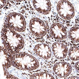
![Immunohistochemistry-Paraffin: Choline kinase alpha Antibody [NBP1-85630] - Staining of human gastrointestinal shows moderate cytoplasmic positivity in glandular cells.](https://images.novusbio.com/fullsize/Choline-kinase-alpha-Antibody-Immunohistochemistry-Paraffin-NBP1-85630-img0014.jpg)
Rabbit Polyclonal
Species Human
Applications WB, IHC, IHC-P
| 1 Publication |
|
![Western Blot: p53 Antibody (PAb 240) [NB200-103] - Analysis of p53 in MCF7 and HeLa lystates. Image courtesy of anonymous customer product review.](https://images.novusbio.com/fullsize/p53-Antibody-PAb-240-Western-Blot-NB200-103-img0001.jpg)
![Immunocytochemistry/Immunofluorescence: p53 Antibody (PAb 240) [NB200-103] - PC12 cells were fixed in 4% paraformaldehyde for 10 minutes and permeabilized in 0.5% Triton X-100 in PBS for 5 minutes. The cells were incubated with anti-p53 Antibody (PAb 240) NB200-103 at 2 ug/ml overnight at 4C and detected with an anti-mouse Dylight 488 (Green) at a 1:1000 dilution for 60 minutes. Nuclei were counterstained with DAPI (Blue). Cells were imaged using a 40X objective.](https://images.novusbio.com/fullsize/p53-Antibody-PAb-240-Immunocytochemistry-Immunofluorescence-NB200-103-img0013.jpg)
Mouse Monoclonal
Species Human, Mouse, Rat
Applications WB, ELISA, Flow
| 3 Reviews 42 Publications |
|
![Western Blot: Fibronectin Antibody - BSA Free [NBP1-91258] - VSOP observed in perivascular-restricted spinal cord lesions with intact BBB. Immunostaining for laminin (brown) shows vascular endothelium and glia limitans of a perivascular lesion, along with infiltrating cells and VSOP (blue). Image collected and cropped by CiteAb from the following publication (https://asn.sagepub.com/lookup/doi/10.1042/AN20120081), licensed under a CC-BY license.](https://images.novusbio.com/fullsize/Fibronectin-Antibody---BSA-Free-Western-Blot-NBP1-91258-img0020.jpg)
![Immunocytochemistry/Immunofluorescence: Fibronectin Antibody - BSA Free [NBP1-91258] - NIH3T3 cells were fixed in 4% paraformaldehyde for 10 minutes and permeabilized in 0.05% Triton X-100 in PBS for 5 minutes. The cells were incubated with anti- NBP1-91258 at 1 ug/ml overnight at 4C and detected with an anti-rabbit Dylight 488 (Green) at a 1:1000 dilution for 60 minutes. Nuclei were counterstained with DAPI (Blue). Cells were imaged using a 100X objective and digitally deconvolved.](https://images.novusbio.com/fullsize/Fibronectin-Antibody---BSA-Free-Immunocytochemistry-Immunofluorescence-NBP1-91258-img0021.jpg)
Rabbit Polyclonal
Species Human, Mouse, Rat
Applications WB, Simple Western, ICC/IF
| 11 Reviews 56 Publications |
|

Goat Polyclonal
Species Rat
Applications WB, IHC
| 3 Reviews 37 Publications |
|


Sheep Polyclonal
Species Human
Applications WB, Simple Western, IHC
| 2 Publications |
|


Goat Polyclonal
Species Human
Applications WB, IHC, ICC
| 1 Review 24 Publications |
|


Species Human
Applications BA
| 3 Reviews 818 Publications |
|
![Western Blot: Cytokeratin 7 Antibody (OV-TL12/30) - Azide and BSA Free [NBP2-47940] - Western Blot Analysis of human HeLa cell lysate using Cytokeratin 7 Antibody (OV-TL12/30).](https://images.novusbio.com/fullsize/Cytokeratin-7-Antibody-OV-TL12-30-Azide-and-BSA-Free-Western-Blot-NBP2-47940-img0007.jpg)
![Immunocytochemistry/Immunofluorescence: Cytokeratin 7 Antibody (OV-TL12/30) - Azide and BSA Free [NBP2-47940] - Immunofluorescent staining of methanol-fixed HeLa cells. Cytokeratin 7 Antibody (OV-TL12/30) followed by goat anti-Mouse IgG-CF488 (Green). The nuclear counterstain is Red Dot.](https://images.novusbio.com/fullsize/Cytokeratin-7-Antibody-OV-TL12-30-Azide-and-BSA-Free-Immunocytochemistry-Immunofluorescence-NBP2-47940-img0006.jpg)
Mouse Monoclonal
Species Human, Mouse
Applications WB, Simple Western, Flow
| 1 Review 1 Publication |
|
![Western Blot: CYLD Antibody [NB100-56461] - Analysis of CYLD in HeLa cell lysate using this antibody.](https://images.novusbio.com/fullsize/CYLD-Antibody-Western-Blot-NB100-56461-img0006.jpg)
![Immunocytochemistry/Immunofluorescence: CYLD Antibody [NB100-56461] - Staining of HeLa cell using control (rabbit Ig) at 5 ug/ml.](https://images.novusbio.com/fullsize/CYLD-Antibody-Immunocytochemistry-Immunofluorescence-NB100-56461-img0003.jpg)
Rabbit Polyclonal
Species Human
Applications WB, Simple Western, ICC/IF
| 1 Publication |
|
![Western Blot: Cytokeratin 20 Antibody (SA35-03) [NBP2-67309] - Analysis of Cytokeratin 20 on CRC cell lysates with Rabbit anti-Cytokeratin 20 antibody at 1/5,000 dilution. Lysates/proteins at 10 ug/Lane. Predicted band size: 48 kDa Observed band size: 48 kDa Exposure time: 30 seconds; 10% SDS-PAGE gel. Proteins were transferred to a PVDF membrane and blocked with 5% NFDM/TBST for 1 hour at room temperature. The primary antibody at 1/5,000 dilution was used in 5% NFDM/TBST at room temperature for 2 hours. Goat Anti-Rabbit IgG - HRP Secondary Antibody at 1:300,000 dilution was used for 1 hour at room temperature.](https://images.novusbio.com/fullsize/Cytokeratin-20-Antibody-SA35-03-Western-Blot-NBP2-67309-img0008.jpg)
![Immunocytochemistry/Immunofluorescence: Cytokeratin 20 Antibody (SA35-03) [NBP2-67309] - Staining Cytokeratin 20 in CRC cells (green). The nuclear counter stain is DAPI (blue). Cells were fixed in paraformaldehyde, permeabilised with 0.25% Triton X100/PBS.](https://images.novusbio.com/fullsize/Cytokeratin-20-Antibody-SA35-03-Immunocytochemistry-Immunofluorescence-NBP2-67309-img0002.jpg)
Rabbit Monoclonal
Species Human, Rat
Applications WB, Flow, ICC/IF
|
|
![Western Blot: Cytokeratin 15 Antibody (ST04-85) [NBP2-67525] - Analysis of Cytokeratin 15 on A431 cell lysates. Proteins were transferred to a PVDF membrane and blocked with 5% BSA in PBS for 1 hour at room temperature. The primary antibody (1/500) was used in 5% BSA at room temperature for 2 hours. Goat Anti-Rabbit IgG - HRP Secondary Antibody at 1:5,000 dilution was used for 1 hour at room temperature.](https://images.novusbio.com/fullsize/Cytokeratin-15-Antibody-ST04-85-Western-Blot-NBP2-67525-img0003.jpg)
![Immunohistochemistry-Paraffin: Cytokeratin 15 Antibody (ST04-85) [NBP2-67525] - Analysis of paraffin-embedded human skin tissue with Rabbit anti-Cytokeratin 15 antibody washed with ddH2O and PBS, and then probed with the primary antibody at 1/400 dilution for 1 hour at room temperature. The detection was performed using an HRP conjugated compact polymer system. DAB was used as the chromogen. Tissues were counterstained with hematoxylin and mounted with DPX.](https://images.novusbio.com/fullsize/Cytokeratin-15-Antibody-ST04-85-Immunohistochemistry-Paraffin-NBP2-67525-img0007.jpg)
Rabbit Monoclonal
Species Human, Mouse, Rat
Applications WB, Flow, ICC/IF
| 4 Publications |
|
Species Human
Applications EnzAct
|
|
![Western Blot: RPE65 Antibody (401.8B11.3D9) - BSA Free [NB100-355] - Subcellular localization of BEST1 and surface Ca2+-dependent Cl- current in patient-derived iPSC-RPEs. Western blots show similar BEST1 expression levels in WT and patient-derived iPSC-RPEs. Each sample was from one cell lysis (BEST1 and beta-actin, RPE65 and CRALBP were on two gels, respectively). Image collected and cropped by CiteAb from the following publication (https://elifesciences.org/articles/29914), licensed under a CC-BY license.](https://images.novusbio.com/fullsize/RPE65-Antibody-401-8B11-3D9-BSA-Free-Western-Blot-NB100-355-img0013.jpg)
![Immunocytochemistry/Immunofluorescence: RPE65 Antibody (401.8B11.3D9) - BSA Free [NB100-355] - Expression of eye-specific markers in the induced eye-like structures induced from lignin-added ES cells. (a) Higher-magnification image of the RPE like structure induced from ESCs after 12-day culture. (b) Immunostaining of eye- like structures. Eye- like structures induced from ESCs after 12-day culture were stained with antibodies against RPE65 (red) and nuclei were stained with DAPI solution (blue). Scale bar: a = 50 um, b,d = 200 um, c,e = 100 um. PLoS One. 2013 Jun 21;8(6):e66376. doi: 10.1371/journal.pone.0066376.](https://images.novusbio.com/fullsize/RPE65-Antibody-401-8B11-3D9-BSA-Free-Immunocytochemistry-Immunofluorescence-NB100-355-img0014.jpg)
Mouse Monoclonal
Species Human, Mouse, Rat
Applications WB, Simple Western, Flow
| 7 Reviews 98 Publications |
|
