| Submit your blog on Floor Plate Development to be featured! |
| Submit your event on Floor Plate Development to be featured! |


Goat Polyclonal
Species Human, Mouse
Applications WB, IHC, ChIP
| 2 Reviews 270 Publications |
|


Goat Polyclonal
Species Human
Applications WB, IHC, ChIP
| 101 Publications |
|


Goat Polyclonal
Species Human, Mouse
Applications WB, ChIP, ICC
| 3 Reviews 102 Publications |
|


Sheep Polyclonal
Species Human
Applications WB, ICC
| 2 Publications |
|

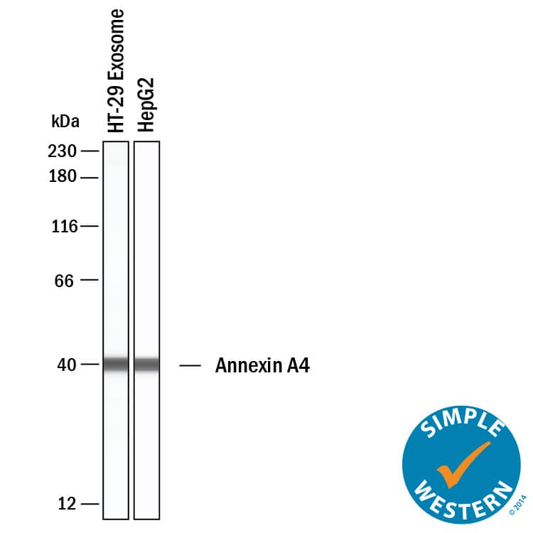
Goat Polyclonal
Species Human, Mouse, Rat
Applications WB, Simple Western, IHC
| 8 Publications |
|

Goat Polyclonal
Species Mouse
Applications WB, IHC
| 2 Reviews 44 Publications |
|


Mouse Monoclonal
Species Human
Applications WB, Flow, CyTOF-ready
| 6 Publications |
|


Species Mouse
Applications BA
| 70 Publications |
|


Mouse Monoclonal
Species Human
Applications Flow, IHC, IP
| 5 Publications |
|
![Western Blot: beta-Galactosidase-1/GLB1 Antibody (OTI1C9) [NBP2-45731] - Analysis of extracts (35ug) from 9 different cell lines by using anti-GLB1 monoclonal antibody (HepG2: human; HeLa: human; SVT2: mouse; A549: human; COS7: monkey; Jurkat: human; MDCK: canine; PC12: rat; MCF7: human).](https://images.novusbio.com/fullsize/beta-Galactosidase-1-GLB1-Antibody-1C9-Western-Blot-NBP2-45731-img0016.jpg)
![Immunohistochemistry: beta-Galactosidase-1/GLB1 Antibody (1C9) [NBP2-45731] - Analysis of Adenocarcinoma of Human ovary tissue. (Heat-induced epitope retrieval by 10mM citric buffer, pH6.0, 120C for 3min)](https://images.novusbio.com/fullsize/beta-Galactosidase-1-GLB1-Antibody-1C9-Immunohistochemistry-NBP2-45731-img0013.jpg)
Mouse Monoclonal
Species Human, Canine, Monkey
Applications WB, IHC, IHC-P
| 6 Publications |
|
![Western Blot: TBX1 Antibody (OTI1C2) [NBP2-46076] - Analysis of HEK293T cells were transfected with the pCMV6-ENTRY control (Left lane) or pCMV6-ENTRY TBX1.](https://images.novusbio.com/fullsize/TBX1-Antibody-1C2-Western-Blot-NBP2-46076-img0002.jpg)
![Immunohistochemistry-Paraffin: TBX1 Antibody (OTI1C2) [NBP2-46076] - Analysis of Human lymph node tissue. (Heat-induced epitope retrieval by 1 mM EDTA in 10mM Tris, pH8.5, 120C for 3min)](https://images.novusbio.com/fullsize/TBX1-Antibody-1C2-Immunohistochemistry-NBP2-46076-img0001.jpg)
Mouse Monoclonal
Species Human, Mouse, Rat
Applications WB, IHC, IHC-P
| 1 Publication |
|
![Immunocytochemistry/Immunofluorescence: TCF15 Antibody [NBP2-55818] - Staining of human cell line HEK 293 shows localization to nuclear speckles. Antibody staining is shown in green.](https://images.novusbio.com/fullsize/TCF15-Antibody-Immunocytochemistry-Immunofluorescence-NBP2-55818-img0001.jpg)
Rabbit Polyclonal
Species Human
Applications ICC/IF
|
|
![Western Blot: SUR1 Antibody (S289-16) [NBP2-59320] - Western Blot analysis of Rat Brain Membrane showing detection of ~160 kDa SUR1 protein using Mouse Anti-SUR1 Monoclonal Antibody, Clone S289-16 (NBP2-59320). Lane 1: Molecular Weight Ladder. Lane 2: Rat Brain Membrane. Load: 15 ug. Block: 2% BSA and 2% Skim Milk in 1X TBST. Primary Antibody: Mouse Anti-SUR1 Monoclonal Antibody (NBP2-59320) at 1:200 for 16 hours at 4C. Secondary Antibody: Goat Anti-Mouse IgG: HRP at 1:1000 for 1 hour RT. Color Development: ECL solution for 6 min in RT. Predicted/Observed Size: ~160 kDa.](https://images.novusbio.com/fullsize/SUR1-Antibody-S289-16-Western-Blot-NBP2-59320-img0007.jpg)
![Immunocytochemistry/Immunofluorescence: SUR1 Antibody (S289-16) [NBP2-59320] - Immunocytochemistry/Immunofluorescence analysis using Mouse Anti-SUR1 Monoclonal Antibody, Clone S289-16 (NBP2-59320). Tissue: Neuroblastoma cells (SH-SY5Y). Species: Human. Fixation: 4% PFA for 15 min. Primary Antibody: Mouse Anti-SUR1 Monoclonal Antibody (NBP2-59320) at 1:50 for overnight at 4C with slow rocking. Secondary Antibody: AlexaFluor 488 at 1:1000 for 1 hour at RT. Counterstain: Phalloidin-iFluor 647 (red) F-Actin stain; Hoechst (blue) nuclear stain at 1:800, 1.6mM for 20 min at RT. (A) Hoechst (blue) nuclear stain. (B) Phalloidin-iFluor 647 (red) F-Actin stain. (C) SUR1 Antibody (D) Composite.](https://images.novusbio.com/fullsize/SUR1-Antibody-S289-16-Immunocytochemistry-Immunofluorescence-NBP2-59320-img0008.jpg)
Mouse Monoclonal
Species Human, Mouse, Rat
Applications WB, ICC/IF, IHC
| 2 Reviews 4 Publications |
|
![Immunohistochemistry: GLI-1 Antibody - BSA Free [NB600-600] - GLI-1 Antibody [NB600-600] - Inhibition of Hh signaling in CAFs blocks its effects on lymphangiogenesis in vivo and in vitro. (E) IHC staining for Ki-67, -SMA, Gli-1 and LYVE1 in tumour sections from mice in the two groups. Human CAFs promote lymphangiogenesis in ovarian cancer via the Hh-VEGF-C signaling axis. Image collected and cropped by CiteAb from the following publication (//pubmed.ncbi.nlm.nih.gov/31012844/) licensed under a CC-BY license.](https://images.novusbio.com/fullsize/GLI-1-Antibody-Immunohistochemistry-Paraffin-NB600-600-img0005.jpg)
![Immunohistochemistry-Paraffin: GLI-1 Antibody [NB600-600] - Analysis of a FFPE mouse brain section using GLI-1 antibody at 1:600 dilution.](https://images.novusbio.com/fullsize/GLI-1-Antibody-Immunohistochemistry-Paraffin-NB600-600-img0003.jpg)
Rabbit Polyclonal
Species Human, Mouse, Canine
Applications WB, ELISA, IB
| 1 Review 57 Publications |
|
![Western Blot: Integrin beta 3/CD61 Antibody (SJ19-09) [NBP2-67416] - Analysis of Integrin beta 3 on different lysates. Proteins were transferred to a PVDF membrane and blocked with 5% BSA in PBS for 1 hour at room temperature. The primary antibody (1/500) was used in 5% BSA at room temperature for 2 hours. Goat Anti-Rabbit IgG - HRP Secondary Antibody at 1:5,000 dilution was used for 1 hour at room temperature. Positive control: Lane 1: HUVEC cell lysateLane 2: mouse marrow tissue lysate](https://images.novusbio.com/fullsize/Integrin-beta-3-CD61-Antibody-SJ19-09-Western-Blot-NBP2-67416-img0012.jpg)
![Immunocytochemistry/Immunofluorescence: Integrin beta 3/CD61 Antibody (SJ19-09) [NBP2-67416] - Staining Integrin beta 3 in HepG2 cells (green). The nuclear counter stain is DAPI (blue). Cells were fixed in paraformaldehyde, permeabilized with 0.25% Triton X-100/PBS.](https://images.novusbio.com/fullsize/Integrin-beta-3-CD61-Antibody-SJ19-09-Immunocytochemistry-Immunofluorescence-NBP2-67416-img0002.jpg)
Rabbit Monoclonal
Species Human, Mouse, Rat
Applications WB, ICC/IF, IHC
| 2 Reviews 7 Publications |
|
