
| Submit your blog on Malignant Odontogenic Tumor to be featured! |
| Submit your event on Malignant Odontogenic Tumor to be featured! |
![Western Blot: Vimentin Antibody [NB300-223] - Analysis of tissue and cell lysates. Antibody at 1:5000 in red. [1] protein standard (red), [2] rat whole brain lysate, [3] HeLa, [4] SH-SY5Y, [5] HEK293, and [6] NIH-3T3 cell lysates. NB300-223 binds to the vimentin protein showing a single band at ~50 kDa. The blot was simultaneously probed with mouse mAb to MAP2C/D, dilution 1:5000 in green, revealing multiple bands around 280 kDa that correspond to full length MAP2A/2B isotypes, and ~70 kDa bands which are MAP2C/D isotypes. MAP2 isotypes are seen only in extracts containing neuronal lineage cells.](https://images.novusbio.com/fullsize/Vimentin-Antibody-Western-Blot-NB300-223-img0002.jpg)
![Immunohistochemistry: Vimentin Antibody [NB300-223] - Attenuated reactive astrocytosis after stroke in Smad1 cKO mice. Enlarged IHC images of boxed areas at the cortical peri-infarct area with the indicated reactive astrocyte markers GFAP, Nestin, and Vimentin. Enlarged images of GFAP IHC highlight the hypertrophic morphology of GFAP+ astrocytes in mutants. Image collected and cropped by CiteAb from the following publication (https://dx.plos.org/10.1371/journal.pone.0136967), licensed under a CC-BY license.](https://images.novusbio.com/fullsize/Vimentin-Antibody-Immunohistochemistry-NB300-223-img0006.jpg)
Chicken Polyclonal
Species Human, Mouse, Rat
Applications WB, ICC/IF, IHC
| 6 Reviews 70 Publications |
|
![Western Blot: UVRAG Antibody [NBP1-18885] - Beclin 1 is acetylated at lysines 430 and 437. TSA and NAM increase the binding of Beclin 1 to Rubicon. Immunoprecipitation of indicated Beclin 1-binding partners with ectopically expressed Flag-Beclin 1 in HEK293T cells treated with TSA and NAM. Image collected and cropped by CiteAb from the following publication (https://www.nature.com/articles/ncomms8215), licensed under a CC-BY license.](https://images.novusbio.com/fullsize/UVRAG-Antibody-Western-Blot-NBP1-18885-img0005.jpg)
![Immunohistochemistry-Paraffin: UVRAG Antibody [NBP1-18885] - Section of human colon carcinoma. Antibody: Affinity purified rabbit anti- UVRAG used at a dilution of 1:200 (1ug/ml). Detection: DAB](https://images.novusbio.com/fullsize/UVRAG-Antibody-Immunohistochemistry-Paraffin-NBP1-18885-img0004.jpg)
Rabbit Polyclonal
Species Human
Applications WB, ICC/IF, IHC
| 3 Publications |
|
![Western Blot: NFkB p105/p50 Antibody (2J10D7) [NB100-56583] - Analysis of p50 in HeLa lysate in the A) absence and B) presence of immunizing peptide using p50 antibody at 5 ug/ml.](https://images.novusbio.com/fullsize/NFkB-p105-p50-Antibody-2J10D7-Western-Blot-NB100-56583-img0004.jpg)
![Immunohistochemistry-Paraffin: NFkB p105/p50 Antibody (2J10D7) [NB100-56583] - IHC-P of rabbit aorta using NB100-56583. Secondary antibody: anti-mouse histofine ( Nisherei Bioscience Inc. ref: 414131F). Development: DAB (Dako ref: K346811-2) and counterstained with Hematoxylin. Submitted via verified customer review](https://images.novusbio.com/fullsize/NFkB-p105-p50-Antibody-2J10D7-Immunohistochemistry-Paraffin-NB100-56583-img0009.jpg)
Mouse Monoclonal
Species Human, Rat, Rabbit
Applications WB, Flow, IHC
| 1 Review 5 Publications |
|
Mouse Monoclonal
Species Human
Applications WB, Simple Western, IHC
|
|
![Simple Western: Cytokeratin 19 Antibody [NB100-687] - Lane view shows a specific band for Cytokeratin 19 in 0.5 mg/ml of HepG2 lysate. This experiment was performed under standard reducing conditions using the 12-230 kDa separation system.](https://images.novusbio.com/fullsize/Cytokeratin-19-Antibody-Simple-Western-NB100-687-img0008.jpg)
![Western Blot: Cytokeratin 19 Antibody [NB100-687] - Western blot detection of YAP and TAZ in proteins extracted from isolated hepatocytes and cholangiocytes and total liver tissue. Antibodies against CK19 and HNF4a were used to confirm purity of the populations and b-actin was used as a loading control. Representative results from a single experiment with n = 3 independent cell isolations and n = 2 total liver tissues. Image collected and cropped by CiteAb from the following publication(https://embomolmed.embopress.org/lookup/doi/10.15252/emmm.201506089), licensed under a CC-BY license.](https://images.novusbio.com/fullsize/Cytokeratin-19-Antibody-Western-Blot-NB100-687-img0010.jpg)
Rabbit Polyclonal
Species Human, Mouse, Rat
Applications WB, Simple Western, Flow
| 1 Review 10 Publications |
|
![Immunocytochemistry/Immunofluorescence: MUC-1 Antibody (SM3) [NB120-22711] - MCF7 cells were fixed for 10 minutes using 10% formalin and then permeabilized for 5 minutes using 1X PBS + 0.05% Triton-X100. The cells were incubated with anti-MUC1 [SM3] at 5 ug/ml overnight at 4C and detected with an anti-mouse IgG Dylight 488 (Green) at a 1:500 dilution. Nuclei were counterstained with DAPI (Blue). Cells were imaged using a 40X objective.](https://images.novusbio.com/fullsize/MUC-1-Antibody-SM3-Immunocytochemistry-Immunofluorescence-NB120-22711-img0004.jpg)
![Immunohistochemistry-Paraffin: MUC-1 Antibody (SM3) [NB120-22711] - IHC analysis of formalin fixed paraffin embedded tissue section of human breast cancer xenograft using MUC-1 antibody clone SM3 at 1:10 dilution. The xenograft section depicted a very specific and intense signal in the periphery of the cancer cells. The necrotic cells also developed a strong immune-positivity while the tumor stroma as well as the nuclei of cells were negative for immunostaining.](https://images.novusbio.com/fullsize/MUC-1-Antibody-SM3-Immunohistochemistry-Paraffin-NB120-22711-img0003.jpg)
Mouse Monoclonal
Species Human, Mouse
Applications ELISA, Flow, ICC/IF
| 14 Publications |
|
![Western Blot: Choline kinase alpha Antibody [NBP1-85630] - Analysis in human cell lines SK-MEL-30 and U-251MG using anti-CHKA antibody. Corresponding CHKA RNA-seq data are presented for the same cell lines. Loading control: anti-HDAC1.](https://images.novusbio.com/fullsize/Choline-kinase-alpha-Antibody-Western-Blot-NBP1-85630-img0008.jpg)
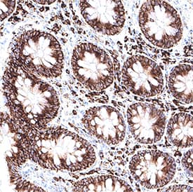
![Immunohistochemistry-Paraffin: Choline kinase alpha Antibody [NBP1-85630] - Staining of human gastrointestinal shows moderate cytoplasmic positivity in glandular cells.](https://images.novusbio.com/fullsize/Choline-kinase-alpha-Antibody-Immunohistochemistry-Paraffin-NBP1-85630-img0014.jpg)
Rabbit Polyclonal
Species Human
Applications WB, IHC, IHC-P
| 1 Publication |
|
![Western Blot: p53 Antibody (PAb 240) [NB200-103] - Analysis of p53 in MCF7 and HeLa lystates. Image courtesy of anonymous customer product review.](https://images.novusbio.com/fullsize/p53-Antibody-PAb-240-Western-Blot-NB200-103-img0001.jpg)
![Immunocytochemistry/Immunofluorescence: p53 Antibody (PAb 240) [NB200-103] - PC12 cells were fixed in 4% paraformaldehyde for 10 minutes and permeabilized in 0.5% Triton X-100 in PBS for 5 minutes. The cells were incubated with anti-p53 Antibody (PAb 240) NB200-103 at 2 ug/ml overnight at 4C and detected with an anti-mouse Dylight 488 (Green) at a 1:1000 dilution for 60 minutes. Nuclei were counterstained with DAPI (Blue). Cells were imaged using a 40X objective.](https://images.novusbio.com/fullsize/p53-Antibody-PAb-240-Immunocytochemistry-Immunofluorescence-NB200-103-img0013.jpg)
Mouse Monoclonal
Species Human, Mouse, Rat
Applications WB, ELISA, Flow
| 3 Reviews 42 Publications |
|
![Immunohistochemistry-Paraffin: S100B Antibody [NBP1-87102] - Analysis in human cerebral cortex and liver tissues. Corresponding S100B RNA-seq data are presented for the same tissues.](https://images.novusbio.com/fullsize/S100B-Antibody-Immunohistochemistry-Paraffin-NBP1-87102-img0020.jpg)
![Western Blot: S100B Antibody [NBP1-87102] - Analysis in mouse cerebral cortex tissue.](https://images.novusbio.com/fullsize/S100B-Antibody-Western-Blot-NBP1-87102-img0010.jpg)
Rabbit Polyclonal
Species Human, Mouse, Rat
Applications WB, ICC/IF, IHC
| 14 Publications |
|
![Western Blot: GRASP65 Antibody (5G8) [NBP2-02665] - Analysis of extracts (35ug) from 9 different cell lines by using anti-GRASP65 monoclonal antibody.](https://images.novusbio.com/fullsize/GRASP65-Antibody-5G8-Western-Blot-NBP2-02665-img0009.jpg)
![Immunocytochemistry/Immunofluorescence: GRASP65 Antibody (OTI5G8) [NBP2-02665] - Staining in Hela cells: red CFTR, Green GRASP65 and Blue is DAPI. Image from a verified customer review.](https://images.novusbio.com/fullsize/GRASP65-Antibody-OTI5G8-Immunocytochemistry-Immunofluorescence-NBP2-02665-img0012.jpg)
Mouse Monoclonal
Species Human, Rat, Porcine
Applications WB, Flow, ICC/IF
| 3 Reviews 2 Publications |
|
![ELISA: Human, Mouse, Rat RelA/NFkB p65 ELISA Kit (Colorimetric) [NBP2-29661] - Standard curve using the internal RelA standard.](https://images.novusbio.com/fullsize/Human-Mouse-Rat-RelA-NFkB-p65-ELISA-Kit-Colorimetric-ELISA-NBP2-29661-img0004.jpg)
![ELISA: Human, Mouse, Rat RelA/NFkB p65 ELISA Kit (Colorimetric) [NBP2-29661] - Nuclear translocation of NF-kB p65 with LPS in Jurkat cells. Jurkat cells, grown to a density of 2X10(6) cells/ml, were treated with LPS (15 ug/ml). The cells were harvested at various indicated time points, and cell lysates were tested in Western analysis using NB100-56712 and also in this ELISA Kit.](https://images.novusbio.com/fullsize/Human-Mouse-Rat-RelA-NFkB-p65-ELISA-Kit-Colorimetric-ELISA-NBP2-29661-img0005.jpg)
Species Human, Mouse, Rat
Applications ELISA
| 95 Publications |
|


Goat Polyclonal
Species Human, Mouse, Rat
Applications WB, Simple Western, IHC
| 2 Reviews 33 Publications |
|
![N/A TIMP-1 [HRP]](https://images.novusbio.com/fullsize2/DATA_TIMP1_DTM100_ELISA_847.jpg)
![N/A TIMP-1 [HRP]](https://images.novusbio.com/fullsize2/TIMP-1_DTM100_ELISA_194.jpg)
Species Human
Applications ELISA
| 97 Publications |
|
![N/A MMP-9 [HRP]](https://images.novusbio.com/fullsize2/DATA_MMP9_DMP900_ELISA_770.jpg)
![N/A MMP-9 [HRP]](https://images.novusbio.com/fullsize2/MMP-9_DMP900_ELISA_148.jpg)
Species Human
Applications ELISA
| 220 Publications |
|
![Simple Western: PCNA Antibody (PC10) [NB500-106] - Image shows a specific band for PCNA in 0.5 mg/mL of HeLa lysate. This experiment was performed under reducing conditions using the 12-230 kDa separation system.](https://images.novusbio.com/fullsize/PCNA-Antibody-PC10-Simple-Western-NB500-106-img0008.jpg)
![Immunocytochemistry/Immunofluorescence: PCNA Antibody (PC10) [NB500-106] - NIH3T3 cells were fixed and permeabilized for 10 minutes using -20C MeOH. The cells were incubated with anti-PCNA Antibody (PC10) NB500-106 at 1 ug/ml overnight at 4C and detected with an anti-mouse Dylight 488 (Green) at a 1:1000 dilution for 60 minutes. Nuclei were counterstained with DAPI (Blue). Cells were imaged using a 100X objective and digitally deconvolved.](https://images.novusbio.com/fullsize/PCNA-Antibody-PC10-Immunocytochemistry-Immunofluorescence-NB500-106-img0015.jpg)
Mouse Monoclonal
Species Human, Mouse, Rat
Applications WB, Simple Western, ChIP
| 5 Reviews 46 Publications |
|
Species Human
Applications EnzAct
|
|
![Western Blot: RPE65 Antibody (401.8B11.3D9) - BSA Free [NB100-355] - Subcellular localization of BEST1 and surface Ca2+-dependent Cl- current in patient-derived iPSC-RPEs. Western blots show similar BEST1 expression levels in WT and patient-derived iPSC-RPEs. Each sample was from one cell lysis (BEST1 and beta-actin, RPE65 and CRALBP were on two gels, respectively). Image collected and cropped by CiteAb from the following publication (https://elifesciences.org/articles/29914), licensed under a CC-BY license.](https://images.novusbio.com/fullsize/RPE65-Antibody-401-8B11-3D9-BSA-Free-Western-Blot-NB100-355-img0013.jpg)
![Immunocytochemistry/Immunofluorescence: RPE65 Antibody (401.8B11.3D9) - BSA Free [NB100-355] - Expression of eye-specific markers in the induced eye-like structures induced from lignin-added ES cells. (a) Higher-magnification image of the RPE like structure induced from ESCs after 12-day culture. (b) Immunostaining of eye- like structures. Eye- like structures induced from ESCs after 12-day culture were stained with antibodies against RPE65 (red) and nuclei were stained with DAPI solution (blue). Scale bar: a = 50 um, b,d = 200 um, c,e = 100 um. PLoS One. 2013 Jun 21;8(6):e66376. doi: 10.1371/journal.pone.0066376.](https://images.novusbio.com/fullsize/RPE65-Antibody-401-8B11-3D9-BSA-Free-Immunocytochemistry-Immunofluorescence-NB100-355-img0014.jpg)
Mouse Monoclonal
Species Human, Mouse, Rat
Applications WB, Simple Western, Flow
| 7 Reviews 98 Publications |
|
