| Submit your blog on Erythropoietic Protoporphyria to be featured! |
| Submit your event on Erythropoietic Protoporphyria to be featured! |
Mouse Monoclonal
Applications IHC
|
|
![Immunocytochemistry/Immunofluorescence: Pancreatic Polypeptide/PP Antibody [NB100-1793] - Histologic comparison of wild type and Seriola dumerili mouse pancreata. Seriola dumerili Ins2 mice (bottom) have normal islet morphology and cyto-architecture compared to littermates with endogenous mouse Ins 1 and Ins 2 (top); pancreatic polypeptide (green; B,E). Scale bar: 100 um. Image collected and cropped by CiteAb from the following publication (www.nature.com/articles/s41598-019-40768-3) licensed under a CC-BY license.](https://images.novusbio.com/fullsize/Pancreatic-Polypeptide-PP-Antibody-Immunocytochemistry-Immunofluorescence-NB100-1793-img0011.jpg)
![Immunohistochemistry-Paraffin: Pancreatic Polypeptide/PP Antibody [NB100-1793] - Staining of paraffin embedded Human Pancreas with antibody at 3 ug/mL. Microwaved antigen retrieval with Tris/EDTA buffer pH 9, HRP-staining.](https://images.novusbio.com/fullsize/Pancreatic-Polypeptide-PP-Antibody-Immunohistochemistry-Paraffin-NB100-1793-img0007.jpg)
Goat Polyclonal
Species Human, Mouse, Rat
Applications Flow, IB, ICC/IF
| 3 Reviews 20 Publications |
|
![Western Blot: Inorganic Pyrophosphatase/PPA1 Antibody [NBP1-31348] - A. 30 ug Neuro2A whole cell lysate/extract. B. 30 ug GL261 whole cell lysate/extract. C. 30 ug C8D30 whole cell lysate/extract. D. 30 ug NIH-3T3 whole cell lysate/extract. E. 30 ug BCL-1 whole cell lysate/extract. F. 30 ug Raw264.7 whole cell lysate/extract. G. 30 ug C2C12 whole cell lysate/extract.](https://images.novusbio.com/fullsize/Inorganic-Pyrophosphatase-PPA1-Antibody-Western-Blot-NBP1-31348-img0012.jpg)
![Immunocytochemistry/Immunofluorescence: Inorganic Pyrophosphatase/PPA1 Antibody [NBP1-31348] - Paraformaldehyde-fixed HeLa, using antibody at 1:200 dilution.](https://images.novusbio.com/fullsize/Inorganic-Pyrophosphatase-PPA1-Antibody-Immunocytochemistry-Immunofluorescence-NBP1-31348-img0008.jpg)
Rabbit Polyclonal
Species Human, Mouse, Rat
Applications WB, ICC/IF, IHC
|
|
![Western Blot: SRPR alpha Antibody [H00006734-B02P] - Analysis of SRPR expression in human pancreas.](https://images.novusbio.com/fullsize/SRPR-alpha-Antibody-Western-Blot-H00006734-B02P-img0002.jpg)
![Immunocytochemistry/Immunofluorescence: SRPR alpha Antibody [H00006734-B02P] - Analysis of purified antibody to SRPR on HeLa cell. (antibody concentration 10 ug/ml)](https://images.novusbio.com/fullsize/SRPR-alpha-Antibody-Immunocytochemistry-Immunofluorescence-H00006734-B02P-img0001.jpg)
Mouse Polyclonal
Species Human
Applications WB, ICC/IF
| 1 Publication |
|
![Immunohistochemistry-Paraffin: Glutaminyl-peptide Cyclotransferase/QPCT Antibody [NBP1-81838] - Staining in human adrenal gland and liver tissues using anti-QPCT antibody. Corresponding QPCT RNA-seq data are presented for the same tissues.](https://images.novusbio.com/fullsize/Glutaminyl-peptide-Cyclotransferase-QPCT-Antibody-Immunohistochemistry-Paraffin-NBP1-81838-img0010.jpg)
![Western Blot: Glutaminyl-peptide Cyclotransferase/QPCT Antibody [NBP1-81838] - Analysis in human cell line SK-MEL-30.](https://images.novusbio.com/fullsize/Glutaminyl-peptide-Cyclotransferase-QPCT-Antibody-Western-Blot-NBP1-81838-img0006.jpg)
Rabbit Polyclonal
Species Human, Mouse
Applications WB, IHC, IHC-P
| 1 Publication |
|
![Western Blot: ALAD Antibody [NBP1-89158] - Lane 1: Marker [kDa] 250, 130, 100, 70, 55, 35, 25, 15, 10. Lane 2: Liver](https://images.novusbio.com/fullsize/ALAD-Antibody-Western-Blot-NBP1-89158-img0004.jpg)
![Immunohistochemistry-Paraffin: ALAD Antibody [NBP1-89158] - Staining in human adrenal gland and pancreas tissues using anti-ALAD antibody. Corresponding ALAD RNA-seq data are presented for the same tissues.](https://images.novusbio.com/fullsize/ALAD-Antibody-Immunohistochemistry-Paraffin-NBP1-89158-img0010.jpg)
Rabbit Polyclonal
Species Human, Rat
Applications WB, Simple Western, IHC
| 1 Publication |
|
![Western Blot: UROD Antibody [NBP2-26190] - Staining of K562 lysate (35 ug protein in RIPA buffer). Primary incubation was 1 hour. Detected by chemiluminescence.](https://images.novusbio.com/fullsize/UROD-Antibody-Western-Blot-NBP2-26190-img0001.jpg)
![Immunohistochemistry-Paraffin: UROD Antibody [NBP2-26190] - (5ug/ml) staining of paraffin embedded Human Kidney. Steamed antigen retrieval with citrate buffer pH 6, AP-staining.](https://images.novusbio.com/fullsize/UROD-Antibody-Immunohistochemistry-Paraffin-NBP2-26190-img0002.jpg)
Goat Polyclonal
Species Human
Applications WB, IHC, IHC-P
| 1 Publication |
|
![Western Blot: FECH Antibody [NBP2-33413] - Lane 1: Marker [kDa] 250, 130, 95, 72, 55, 36, 28, 17, 10. Lane 2: Human cell line RT-4. Lane 3: Human cell line U-251MG](https://images.novusbio.com/fullsize/FECH-Antibody-Western-Blot-NBP2-33413-img0004.jpg)
![Immunohistochemistry-Paraffin: FECH Antibody [NBP2-33413] - Staining of human lymph node shows no positivity in germinal center cells as expected.](https://images.novusbio.com/fullsize/FECH-Antibody-Immunohistochemistry-Paraffin-NBP2-33413-img0007.jpg)
Rabbit Polyclonal
Species Human
Applications WB, IHC, IHC-P
| 1 Publication |
|
![Immunohistochemistry-Paraffin: HMBS Antibody [NBP2-33600] - Staining in human bone marrow and cerebral cortex tissues using anti-HMBS antibody. Corresponding HMBS RNA-seq data are presented for the same tissues.](https://images.novusbio.com/fullsize/HMBS-Antibody-Immunohistochemistry-Paraffin-NBP2-33600-img0012.jpg)
![Western Blot: HMBS Antibody [NBP2-33600] - Lane 1: Marker [kDa] 250, 130, 95, 72, 55, 36, 28, 17, 10<br/>Lane 2: Human cell line RT-4](https://images.novusbio.com/fullsize/HMBS-Antibody-Western-Blot-NBP2-33600-img0003.jpg)
Rabbit Polyclonal
Species Human, Mouse
Applications WB, ICC/IF, IHC
| 2 Publications |
|


Goat Polyclonal
Species Human, Mouse, Rat
Applications WB, Simple Western, ICC
| 3 Reviews 15 Publications |
|


Mouse Monoclonal
Species Human
Applications WB, Simple Western, IHC
| 3 Reviews 56 Publications |
|
Species Human
Applications BA
| 51 Publications |
|


Species Human
Applications BA
| 21 Publications |
|
![Immunohistochemistry-Paraffin: Desmoplakin Antibody [NBP2-48836] - Staining in human skin and skeletal muscle tissues using anti-DSP antibody. Corresponding DSP RNA-seq data are presented for the same tissues.](https://images.novusbio.com/fullsize/Desmoplakin-Antibody-Immunohistochemistry-Paraffin-NBP2-48836-img0005.jpg)
![Immunohistochemistry-Paraffin: Desmoplakin Antibody [NBP2-48836] - Staining of human skeletal muscle shows low expression as expected.](https://images.novusbio.com/fullsize/Desmoplakin-Antibody-Immunohistochemistry-Paraffin-NBP2-48836-img0003.jpg)
Rabbit Polyclonal
Species Human, Canine
Applications ICC/IF, IHC, IHC-P
| 2 Publications |
|
![Western Blot: Acetylcholinesterase/ACHE Antibody [NB100-1519] - Staining (0.3ug/ml) of Jurkat (A) and (0.5ug/ml) HepG2 (B) cell lysate (35ug protein in RIPA buffer). Detected by chemiluminescence.](https://images.novusbio.com/fullsize/Acetylcholinesterase-ACHE-Antibody-Western-Blot-NB100-1519-img0006.jpg)
![Immunocytochemistry/Immunofluorescence: Acetylcholinesterase/ACHE Antibody [NB100-1519] - Immunofluorescence analysis of paraformaldehyde fixed U2OS cells, permeabilized with 0.15% Triton. Primary incubation 1hr (10ug/ml) followed by Alexa Fluor 488 secondary antibody (2ug/ml), showing nuclear, membrane and cytoplasmic staining. The nuclear stain is DAPI (blue). Negative control: Unimmunized goat IgG (10ug/ml) followed by Alexa Fluor 488 secondary antibody (2ug/ml).](https://images.novusbio.com/fullsize/Acetylcholinesterase-ACHE-Antibody-Immunocytochemistry-Immunofluorescence-NB100-1519-img0005.jpg)
Goat Polyclonal
Species Human, Mouse, Rat
Applications WB, Flow, ICC/IF
| 1 Review 1 Publication |
|
![Western Blot: ALAS2 Antibody (6C1) [H00000212-M01] - Analysis of ALAS2 expression in transfected 293T cell line by ALAS2 monoclonal antibody (M01), clone 6C1.Lane 1: ALAS2 transfected lysate(64 KDa).Lane 2: Non-transfected lysate.](https://images.novusbio.com/fullsize/ALAS2-Antibody-6C1-Western-Blot-H00000212-M01-img0006.jpg)
![Western Blot: ALAS2 Antibody (6C1) [H00000212-M01] - ALAS2 monoclonal antibody (M01), clone 6C1. Analysis of ALAS2 expression in Raw 264.7.](https://images.novusbio.com/fullsize/ALAS2-Antibody-6C1-Western-Blot-H00000212-M01-img0005.jpg)
Mouse Monoclonal
Species Human, Mouse
Applications WB, ELISA
| 4 Publications |
|
![Western Blot: CPOX Antibody (36B10) [NBP2-59438] - Lane 1: Recombinant protein CPOX, Lane 2: 293T cell lysates, Lane 3: HepG2 cell lysates, Lane 4: A549 cell lysates, Lane 5: Jurkat cell lysates, Lane 6: K562 cell lysates, Lane 7: LnCaP cell lysates](https://images.novusbio.com/fullsize/CPOX-Antibody-36B10-Western-Blot-NBP2-59438-img0001.jpg)
![Immunocytochemistry/Immunofluorescence: CPOX Antibody (36B10) [NBP2-59438] - Analysis of CPOX in Hep3B cells line, stained with DAPI (Blue) for nucleus staining and monoclonal anti-human CPOX antibody (1:100) with goat anti-mouse IgG-Alexa fluor 488 conjugate (Green).](https://images.novusbio.com/fullsize/CPOX-Antibody-36B10-Immunocytochemistry-Immunofluorescence-NBP2-59438-img0002.jpg)
mouse Monoclonal
Species Human
Applications WB, ELISA, ICC/IF
| 1 Publication |
|
![Western Blot: PPOX Antibody (2F10) [H00005498-M01] - Analysis of PPOX expression in transfected 293T cell line by PPOX monoclonal antibody (M01), clone 2F10.Lane 1: PPOX transfected lysate(50.8 KDa).Lane 2: Non-transfected lysate.](https://images.novusbio.com/fullsize/PPOX-Antibody-2F10-Western-Blot-H00005498-M01-img0009.jpg)
![Immunohistochemistry-Paraffin: PPOX Antibody (2F10) [H00005498-M01] - Analysis of monoclonal antibody to PPOX on formalin-fixed paraffin-embedded human lung, adenosquamous cell carcinoma. Antibody concentration 3 ug/ml.](https://images.novusbio.com/fullsize/PPOX-Antibody-2F10-Immunohistochemistry-Paraffin-H00005498-M01-img0006.jpg)
Mouse Monoclonal
Species Human, Mouse
Applications WB, ELISA, IHC
| 3 Publications |
|
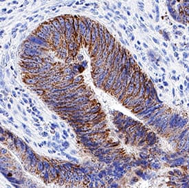
![Immunohistochemistry-Paraffin: EPX Antibody (rEPO104) [NBP3-07388] - Formalin-fixed, paraffin-embedded human Pancreas stained with EPX Recombinant Mouse Monoclonal Antibody (rEPO104).](https://images.novusbio.com/fullsize/EPX-Antibody-rEPO104-Immunohistochemistry-Paraffin-NBP3-07388-img0001.jpg)
Mouse Monoclonal
Species Human
Applications Flow, ICC/IF, IHC
|
|
![Immunohistochemistry-Paraffin: POMT1 Antibody [NBP3-12487] - 1:100 dilution in IHC blocking buffer. DAB (brown) staining and Hematoxylin QS (blue) counterstain. 40x magnification.](https://images.novusbio.com/fullsize/POMT1-Antibody-Immunohistochemistry-Paraffin-NBP3-12487-img0001.jpg)
Rabbit Polyclonal
Species Human, Mouse, Rat
Applications WB, ELISA, IHC
|
|
